Fusion imaging gene and its lentiviral expression plasmid, lentivirus, cell, its preparation method and application

A technology for expressing plasmids and lentiviruses, which can be applied to cells and can solve the problems of successful transfection of difficult lentiviral particles, increasing the length of foreign genes, and inactivating functions.

Active Publication Date: 2022-03-11
ACADEMY OF MILITARY MEDICAL SCI
View PDF4 Cites 0 Cited by
  • Summary
  • Abstract
  • Description
  • Claims
  • Application Information

AI Technical Summary

Problems solved by technology

Lentivirus is the most commonly used carrier for stable gene transfection labeling, but there are no reports on lentiviral particles carrying multimodal imaging genes (quantitative imaging, morphological imaging, functional imaging) and their transfected labeled cells. There are many technical difficulties in the above purpose, including: 1) The length of multiple reporter genes is relatively large, and the general lentiviral vector is not easy to accommodate, and it is not easy to successfully transfect the target cells
In particular, the use of multiple promoters to drive the expression of each reporter gene separately will further increase the length of the foreign gene, making it more difficult to achieve effective packaging of lentiviral particles and successful transfection of target cells; 2) Linking multiple reporter genes to form a fusion For multimodal imaging genes, the use of a promoter to drive expression can save the length occupied by the promoter, but inappropriate fusion can lead to functional inactivation due to spatial interaction between individual gene products
In addition, the fused gene may also be difficult to obtain highly active lentivirus due to its large length

Method used


Image

  • Fusion imaging gene and its lentiviral expression plasmid, lentivirus, cell, its preparation method and application
  • Fusion imaging gene and its lentiviral expression plasmid, lentivirus, cell, its preparation method and application
  • Fusion imaging gene and its lentiviral expression plasmid, lentivirus, cell, its preparation method and application

Examples


preparation example Construction

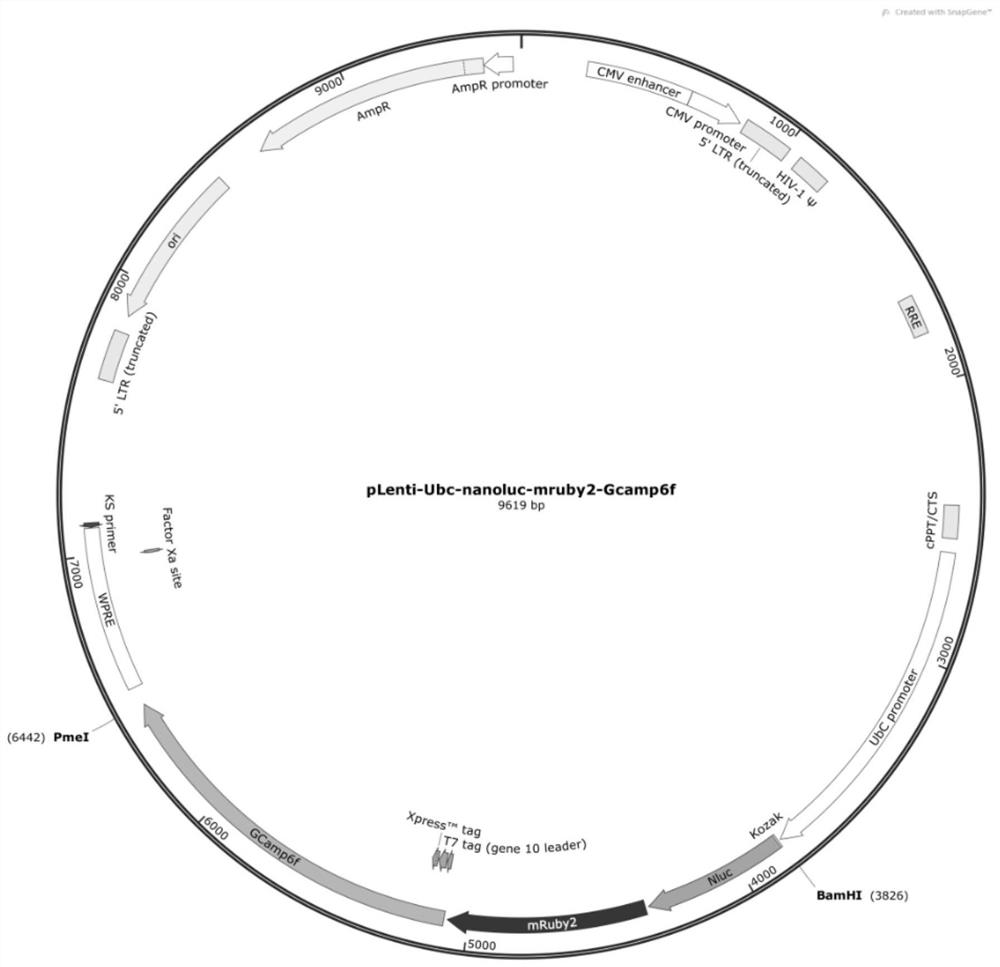
[0093] The sixth aspect of the embodiment of the present invention relates to the preparation method of the above-mentioned lentivirus. The above-mentioned pLenti-Ubc-Nanoluc-mRuby2-Gcamp6f plasmid and the lentivirus packaging plasmid are co-transfected into cells, and the obtained lentivirus carrying the fusion imaging gene is packaged. Include at least the following steps:

[0094] Co-transfect 293T cells with pLenti-Ubc-Nanoluc-mRuby2-Gcamp6f plasmid and lentiviral packaging plasmids PAX2 and pMD2G to obtain lentiviral particles carrying fusion imaging genes;

[0095] Preferably, the mass ratio of the pLenti-Ubc-Nanoluc-mRuby2-Gcamp6f plasmid, the plasmid PAX2, and the plasmid pMD2G3 is 3:2:1.

[0096] The seventh aspect of the embodiment of the present invention relates to the above-mentioned fused imaging gene-marked cells or cells infected by the above-mentioned lentivirus, preferably, the cells are selected from human H1 embryonic stem cells and human pluripotent stem c...

Embodiment 1

[0098] Example 1: Preparation of lentiviral particles carrying fusion multimodal imaging genes

[0099] (1) Design and construction of fused multimodal imaging genes

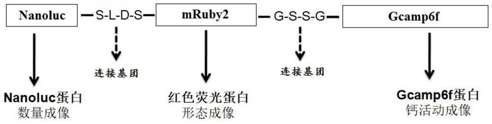
[0100] 1) In order to reduce the impact of fusion expression of multiple imaging genes on protein folding and spatial conformation, and maintain the activity of each protein, the flexible polypeptide sequences S-L-D-S (SEQ ID NO.8) and G-S-S-G (SEQ ID NO.8) and G-S-S-G (SEQ ID NO. ID NO.9) as a linking group (Linker) for connection, such as figure 1 The multi-gene fusion expression design structure diagram shown;

[0101] 2) Obtain Nanoluc gene and mRuby2 gene by gene synthesis, remove the tail stop codon, add BamHI restriction sequence (GGATCC) to the front of Nanoluc gene, and add an S amino acid gene sequence (TCG) and Xhol restriction sequence (CTCGAG) at the end ; Add a SalI restriction sequence (GTCGAC, Xhol isotail enzyme) and an S amino acid (TCG) to the front end of mRuby2 successively; add a G amino ...

Embodiment 2

[0129] Example 2: Application of fused multimodal imaging genes in human pluripotent stem cells

[0130] (1) Multimodal Imaging Gene Markers of Human Embryonic Stem Cells (hESCs)

[0131] Through the conventional lentivirus infection cell method (refer to PLoS One.2013; 8(6):e66369.), use the above-mentioned lentiviral particles carrying fusion gene probes to transfect h1ESC (gifted by Professor Cao Nan of Sun Yat-Sen University, or from ATCC, USA purchased), and the positive clones were isolated and amplified by a patented method (ZL201911133148.1). Experimental results such as Figure 8 shown by Figure 8 It can be seen that the lentiviral particles carrying the fused multimodal imaging gene prepared by the present invention successfully transfect hESC, the expression of mRuby2 can be observed, and the hESC cell line fused with the imaging gene marker is successfully obtained. It is suggested that the lentiviral particles carrying multimodal imaging genes prepared by the ...


PUM


Abstract

The invention relates to the field of biotechnology, in particular to a fusion imaging gene and its lentiviral expression plasmid, lentivirus and cells, its preparation method and application. The fusion imaging gene includes a bioluminescence imaging gene, a fluorescent protein gene and a calcium imaging gene, and the three genes are connected through a linking group. Insert the fusion gene into the transformed lentiviral expression plasmid to obtain lentiviral particles carrying the fusion imaging gene. Lentiviral particles can be used to infect different types of cells to obtain fusion imaging gene-labeled cells. Cells marked by the fusion imaging gene in the present invention can simultaneously perform cell number imaging detection, morphological imaging detection and calcium activity functional imaging detection, which is of great use in cell fate tracking.

Description

technical field [0001] The invention relates to the field of biotechnology, in particular to a fusion imaging gene and its lentiviral expression plasmid, lentivirus, cell, its preparation method and application. Background technique [0002] Imaging tracing of cell fate has extensive and important applications in the field of biomedical research, such as evaluation of cell responses to specific stimuli, evaluation of cells after transplantation, and so on. Taking cell transplantation research as an example, cell fate tracing, including survival, distribution, differentiation, and functional integration, is an important indicator for evaluating its safety and effectiveness. In previous studies, due to the lack of multimodal imaging gene-marked cell lines, simultaneous tracking of cell survival / distribution, differentiation / morphology, and functional integration after transplantation could not be achieved in the same cell. In order to achieve the above-mentioned different tra...

Claims


Application Information

Patent Timeline
Patent Type & Authority Patents(China)
IPC IPC(8): C12N15/62C12N15/867C12N15/66C12N15/65C12N5/10
CPCC07K14/47C12N15/86C12N15/65C12N5/0686C12N5/0606C12N5/0696C07K2319/60C07K2319/61C12N2740/15043C12N2800/107C12N2510/00
Inventor 刘志强袁增强
Owner ACADEMY OF MILITARY MEDICAL SCI
Why Eureka
Try Eureka