
Thrombin detection method based on magnetic separation of deoxyribozyme and circular cutting and thrombin kit

A deoxyribozyme and detection method technology, applied in the field of thrombin detection, can solve the problems of lost samples, difficulty in maintaining the activity of biological macromolecules, etc., and achieve the effects of improving accuracy, convenient calculation, and reducing interference

Active Publication Date: 2021-11-23
WEST CHINA HOSPITAL SICHUAN UNIV
View PDF8 Cites 3 Cited by
  • Summary
  • Abstract
  • Description
  • Claims
  • Application Information

AI Technical Summary

Problems solved by technology

Precipitation and centrifugation are difficult to maintain the activity of biomacromolecules and may lose sample or co-centrifuge with unwanted impurities and precipitate

Method used

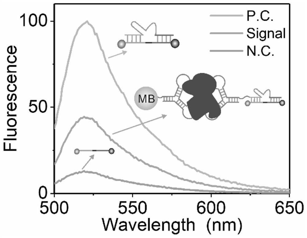

Image

  • Thrombin detection method based on magnetic separation of deoxyribozyme and circular cutting and thrombin kit
  • Thrombin detection method based on magnetic separation of deoxyribozyme and circular cutting and thrombin kit
  • Thrombin detection method based on magnetic separation of deoxyribozyme and circular cutting and thrombin kit

Examples


Embodiment 1

[0026] A thrombin detection method based on magnetic separation of deoxyribozyme and cyclic cutting, comprising the following steps:

[0027] S1 Preparation of magnetic bead dispersion: Take 50 mL of magnetic beads with a concentration of 10 mg / mL, add them to a 600 μL centrifuge tube, then add 200 μL of coupling buffer solution and mix well, shake at 1800 rpm for 20 minutes at 37 °C, and separate the supernatant;

[0028] S2 washing: add 100 μL of coupling buffer solution, mix well, shake at 1800 rpm for 20 min at 37°C, separate the supernatant, and repeat 2 times;

[0029] S3 coupling: add 295 μL of coupling buffer solution to the magnetic beads, add 4 μL of 100 μM biotin-modified thrombin aptamer DNA stock solution (abbreviated as P-L in English), and the biotin-modified thrombin aptamer core The nucleotide sequence is shown in SEQ ID NO.1, specifically: TTTTTTTTTTTTTTTTTTTTTTTTTTTTTTTTTTTAGTCCGTGGTAGGGCAGGTTGGGGTGACT), the 5' end is modified with biotin (Biotin), mixed, sh...

Embodiment 2

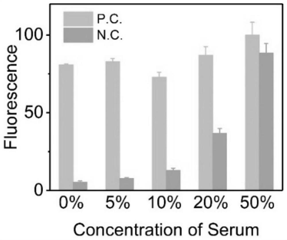
[0034] Using the method of Example 1, the difference from Example 1 is that 0%, 5%, 10%, 20% and 50% of the serum in step S4 are respectively replaced by 5 μL of thrombin (0.01% Tween-20, 30% glycerol, 1×PBS), the results are as follows figure 2 as shown, figure 2 Among them, P.C. is the signal group, and N.C. is the background group. figure 2 It can be seen that the difference in fluorescence intensity between the signal group and the background group is getting smaller and smaller, but there is still a significant difference in fluorescence intensity at 50%, so the anti-interference ability is strong, and thrombin can be detected in 10% serum.

Embodiment 3

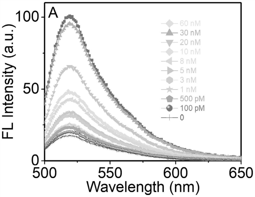
[0036] Adopt the method of embodiment 1, but the concentration of thrombin is respectively 0, 100pM, 500pM, 1nM, 3nM, 5nM, 8nM, 10nM, 20nM, 30nM and 60nM, detection result is as follows image 3 as shown, image 3 The curves from bottom to top correspond to 0, 100pM, 500pM, 1nM, 3nM, 5nM, 8nM, 10nM, 20nM, 30nM, and 60nM, which are superimposed on the top curve. image 3It can be seen that when the wavelength is around 525nm, there is also fluorescence intensity when the concentration of thrombin is 100pM, and the fluorescence intensity value is relatively high, so the detection limit is high and the detection limit reaches 100pM;

[0037] Fit the concentration of thrombin with the fluorescence intensity, the result is as follows Figure 4 shown by Figure 4 It can be seen that when the thrombin concentration is between 100pM-30nM, the relationship between the fluorescence intensity and the thrombin concentration is linear, the linear equation is y=2.42x+21.3, and the correla...


PUM

PropertyMeasurementUnit
concentrationaaaaaaaaaa

Abstract

The invention discloses a thrombin detection method based on magnetic separation of deoxyribozyme and cyclic cleavage and a thrombin kit, and the thrombin detection method comprises the following steps: adding magnetic beads into a coupling buffer solution, uniformly mixing, centrifugally separating the supernatant, washing, adding a coupling buffer solution and a DNA stock solution of thrombin modified with biotin, centrifugally separating a supernatant, and washing with an incubation buffer solution for 2-3 times to obtain a magnetic bead dispersion liquid, wherein the magnetic bead concentration in the magnetic bead dispersion liquid is 4-6mg / mL; uniformly mixing the magnetic bead dispersion liquid, thrombin, a thrombin aptamer modified with deoxyribozyme and an incubation buffer solution, separating a supernatant, washing the supernatant, and dissolving the obtained solid in an enzyme digestion buffer solution to obtain a mixed solution; and adding the mixed solution into an enzyme digestion buffer solution containing a substrate chain, oscillating and incubating for 2.5-3.5 hours, carrying out magnetic separation, taking supernate, carrying out fluorescence detection, and judging the content of thrombin according to the detected fluorescence intensity. The anti-interference capability is strong, and the sensitivity is high.

Description

technical field [0001] The invention belongs to the technical field of thrombin detection, and in particular relates to a thrombin detection method and kit based on magnetic separation of deoxyribozymes and cyclic cutting. Background technique [0002] Thrombin usually exists in blood or serum, and the matrix is ​​usually relatively complex, so the matrix interference of biological samples is particularly serious. Therefore, when analyzing trace target analytes in biological matrices, the original sample must be separated and purified; after separation and purification, it is tested by conventional detection methods, including electrophoresis, centrifugation, ultrafiltration and precipitation, etc. . Electrophoresis is inexpensive, but time-consuming and not reproducible. Ultrafiltration has high separation efficiency, but may adsorb biomacromolecules. Precipitation and centrifugation methods are difficult to maintain the activity of biomolecules, and may lose samples or ...

Claims


Application Information

Patent Timeline
Patent Type & Authority Applications(China)
IPC IPC(8): G01N33/573G01N21/64
CPCG01N33/573G01N21/6486G01N2333/974
Inventor 程南生樊玮陈骏伯杨鹏胡昌佳
Owner WEST CHINA HOSPITAL SICHUAN UNIV
Learn More