Contrast solution for the characterisation of biological samples by electron or correlative microscopy
A solution and general formula technology, applied in the preparation of test samples, sampling, microlithography exposure equipment, etc.
- Summary
- Abstract
- Description
- Claims
- Application Information
AI Technical Summary
Problems solved by technology
Method used
Image

Examples
Embodiment 1
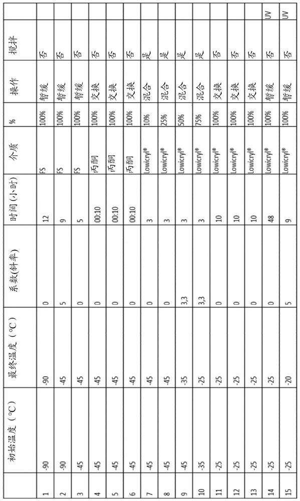
[0062] PTA-YbCl 3 Preparation of solutions (stock solutions)
[0063] A solution of phosphotungstic acid (concentration 3.2 mM) in 10 ml of water / ethanol containing 20% (v / v) ethanol was prepared. The pH of the solution was brought to about 5 with sodium hydroxide 1M. To this solution was added an equal volume of a solution of ytterbium chloride hexahydrate 48 mM (final concentration) in ethanol / water containing 20% (v / v) ethanol. The pH was adjusted again to 5 with sodium hydroxide 1M. The mixture was kept under stirring at room temperature overnight. The solution thus obtained was called "X solution 1.5" and showed a precipitate. The precipitate was removed by filtration, and the pH was adjusted again to about 5 by adding a 20 mM solution of MES. The solution was then again kept under stirring overnight and filtered again, this time through a membrane with a pore size of 200 nm, in order to obtain a solution free of precipitates and referred to as "stock solution"...
Embodiment 2
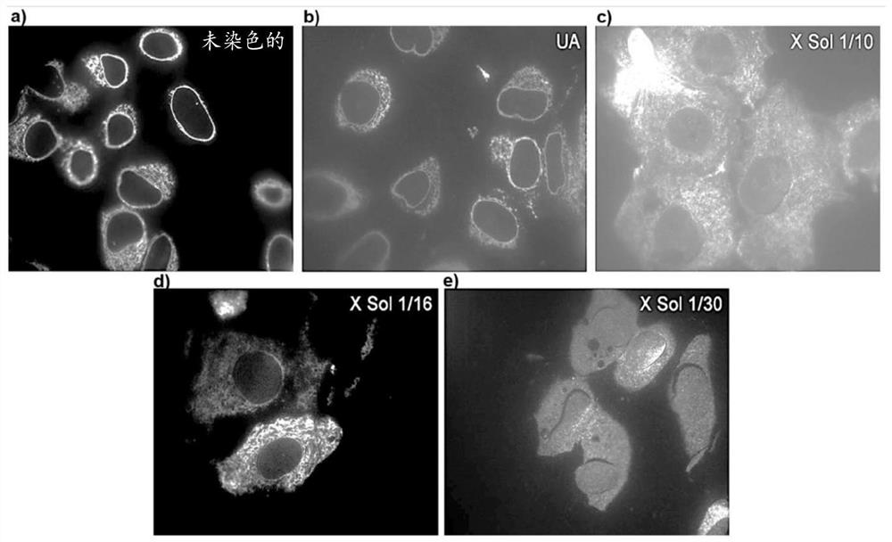
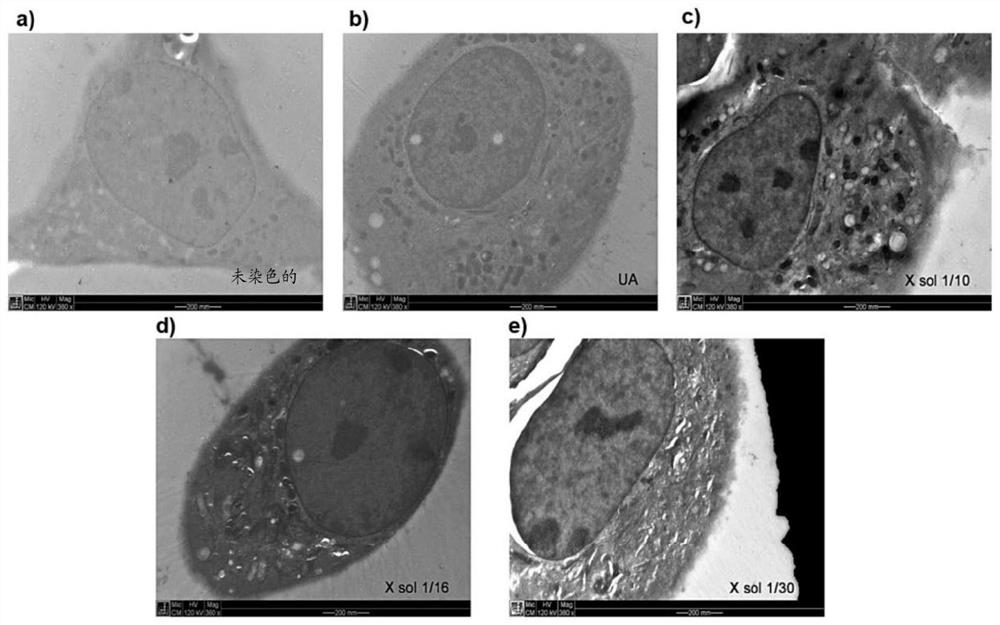
[0065] Use of the solution according to the invention as a contrast medium for correlative microscopy using the HPF / FS protocol (CLEM) Characterization of Biological Samples
[0066] The stock solution (0.2 ml) obtained according to Example 1 was lyophilized and dissolved in 2 ml ("X Sol 1 / 10"), 3.2 ml ("X Sol 1 / 16") or 6 ml ("X Sol 1 / 30") ) in a water / acetone mixture containing 95% (v / v) acetone in order to obtain the solutions according to the invention in three different concentrations respectively.
[0067] Each solution was centrifuged, and the supernatant was used on a cell line stably transfected with a fluorescent protein conjugated to the endoplasmic reticulum in the standard HPF / FS protocol ( figure 1 medium in the diagram). These were used as standard samples for correlation protocols. Cells embedded in this protocol are processed by high pressure freezing / water displacement (HPF / FS) with organic solvents at cryogenic temperatures. Sample sections (cut by us...
Embodiment 3
[0071] Use of the solution according to the invention as a contrast agent for use in HPF / FS protocols by correlative microscopy (CLEM) Characterization of biological samples (Drosophila oocytes)
[0072] Experiments similar to those described in Example 2 were performed using Drosophila oocytes as biological samples and ethanol as the organic solvent instead of acetone. The images obtained are in Figure 5 shown in .
[0073] The images thus obtained show that the solutions of the invention are able to obtain comparable results in terms of fluorescence emission, while at the same time ensuring a better contrast than when using a solution comprising uranyl acetate, thus demonstrating that after correlation microscopy ( CLEM) characterization is more effective.
PUM
 Abstract
Description
Claims
Application Information
 - R&D
- Intellectual Property
- Life Sciences
- Materials
- Tech Scout
- Unparalleled Data Quality
- Higher Quality Content
- 60% Fewer Hallucinations
Browse by: Latest US Patents, China's latest patents, Technical Efficacy Thesaurus, Application Domain, Technology Topic, Popular Technical Reports.
© 2025 PatSnap. All rights reserved.Legal|Privacy policy|Modern Slavery Act Transparency Statement|Sitemap|About US| Contact US: help@patsnap.com