Genetic coding nanoprobe for cell membrane potential detection and preparation method and application thereof

A potential detection and nano-probe technology, applied in the fields of genetic engineering and brain science, to achieve specific labeling and cell excitability sensing, enhance spatial penetration and time resolution

Pending Publication Date: 2022-01-28
SUZHOU INST OF NANO TECH & NANO BIONICS CHINESE ACEDEMY OF SCI
View PDF10 Cites 0 Cited by
  • Summary
  • Abstract
  • Description
  • Claims
  • Application Information

AI Technical Summary

Problems solved by technology

The research on the detection of cell membrane potential by optical genetic means combined with nanoprobes is still seldom carried out.

Method used


Image

  • Genetic coding nanoprobe for cell membrane potential detection and preparation method and application thereof
  • Genetic coding nanoprobe for cell membrane potential detection and preparation method and application thereof
  • Genetic coding nanoprobe for cell membrane potential detection and preparation method and application thereof

Examples


preparation example Construction

[0065] Another aspect of the embodiments of the present invention also provides a method for preparing the aforementioned genetically encoded nanoprobe for cell membrane potential detection, which includes:

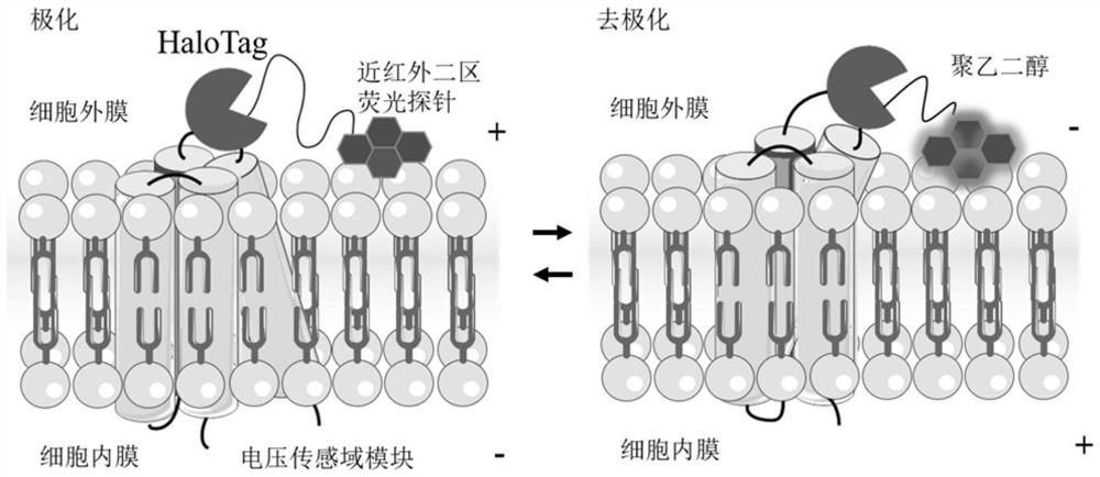
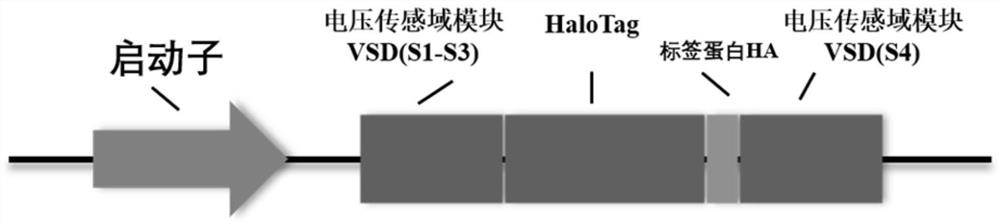
[0066] Construct an adenovirus containing a reporter group, and then infect neuron cells to obtain neurons expressing the recombinant protein; the reporter gene includes the voltage sensing domain VSD gene inserted into the module and the HaloTag gene inserted into the module tag protein;

[0067] Linking the HaloTag ligand and linker with the fluorescent probe in the second near-infrared region through a reaction; forming a fluorescent probe in the second region of near-infrared; and,

[0068] Dilute and disperse the obtained near-infrared second region fluorescent probe in a buffer solution, incubate neurons expressing the recombinant protein, realize the specific labeling of the near infrared second region fluorescent probe and neurons, and obtain a genetically encoded ...

Embodiment 1

[0092] Example 1 Neuron Cell Membrane Potential Detection Method

[0093] 1. Optical genetic tools to build neuron models

[0094] Create neurons carrying the voltage-sensing domain VSD and the tag protein HaloTag gene:

[0095] The sequence of the voltage sensing domain VSD gene used is shown in the aforementioned SEQ ID No. 1, and the sequence of the tag protein HaloTag gene is shown in the aforementioned SEQ ID No. 2.

[0096] 1.1 Preparation of adenovirus carrying HaloTag gene

[0097] Design and insert the gene containing VSD-HaloTag into the pTrack plasmid, digest and linearize the shuttle vector containing the recombinant protein vector, recombine the shuttle vector containing the target gene and the backbone vector pAdeasy in the competent strain BJ5183, and obtain the pAdeasy containing VSD-HaloTag plasmid;

[0098] 1.2 Lipofectamine transfection and adenovirus expansion

[0099] Mix the pAdeasy plasmid containing VSD-HaloTag with liposomes, add the liposome-plasmid...


PUM

PropertyMeasurementUnit
wavelengthaaaaaaaaaa

Abstract

The invention discloses a genetic coding nanoprobe for cell membrane potential detection and a preparation method and an application thereof. The genetic coding nanoprobe comprises an optical genetic module which is mainly composed of an adenovirus and neurons infected by the adenovirus and is expressed on a neuron cell membrane infected by an adenovirus gene, reporter genes of the optical genetic module comprise an inserted module voltage sensing domain VSD gene and an inserted module tag protein HaloTag gene, and the sequences of the VSD gene and the HaloTag gene are respectively as shown in SEQ ID No. 1 and SEQ ID No. 2; and a near-infrared two-region fluorescent probe mainly comprises a near-infrared two-region organic nano probe, a quantum dot probe and a length-adjustable connexon. According to the invention, a genetic coding membrane potential detection method is cooperated with a near-infrared two-region fluorescence technology, so that the electrophysiological activity of a single neuron can be observed, and a method is provided for observing the electrical activity of a neuron cell membrane.

Description

technical field [0001] The invention relates to a nano-probe, in particular to a genetically coded near-infrared second region nano-probe for cell membrane potential detection and a preparation method and application thereof, belonging to the technical fields of genetic engineering and brain science. Background technique [0002] One of the research focuses of brain science is the structure and function of the brain. Real-time observation of neuron activity and structural tracking of specific neural circuits at the cellular level are essential for analyzing the structure and function of the brain. At present, the neurobiological observation methods relying on the two basic physical signals of light and electricity mainly include microelectrode detection, calcium imaging, and voltage-sensitive imaging. In voltage-sensitive imaging, fluorescence imaging uses chemical probes, namely voltage-sensitive dyes, to infuse targeted cells or tissues. When the excitation light is irradi...

Claims


Application Information

Patent Timeline
Patent Type & Authority Applications(China)
IPC IPC(8): C12N15/861C12N15/65C09K11/06G01N21/64
CPCC12N15/86C12N15/65C09K11/06G01N21/6486C12N2710/10343
Inventor 陈光村鲁雅昕王强斌刘永阳
Owner SUZHOU INST OF NANO TECH & NANO BIONICS CHINESE ACEDEMY OF SCI
Why Eureka
Try Eureka