Imaging-based pooled crispr screening

A reporter gene and sequence technology, applied in the field of cell imaging, can solve problems such as difficulty in determining the genotype of a single cell

Pending Publication Date: 2022-01-28
PRESIDENT & FELLOWS OF HARVARD COLLEGE
View PDF12 Cites 0 Cited by
  • Summary
  • Abstract
  • Description
  • Claims
  • Application Information

AI Technical Summary

Problems solved by technology

Despite imaging's ability to obtain cellular phenotypes, imaging-based pooled library screening remains challenging, primarily b...

Method used


Image

  • Imaging-based pooled crispr screening
  • Imaging-based pooled crispr screening
  • Imaging-based pooled crispr screening

Examples


Embodiment 1

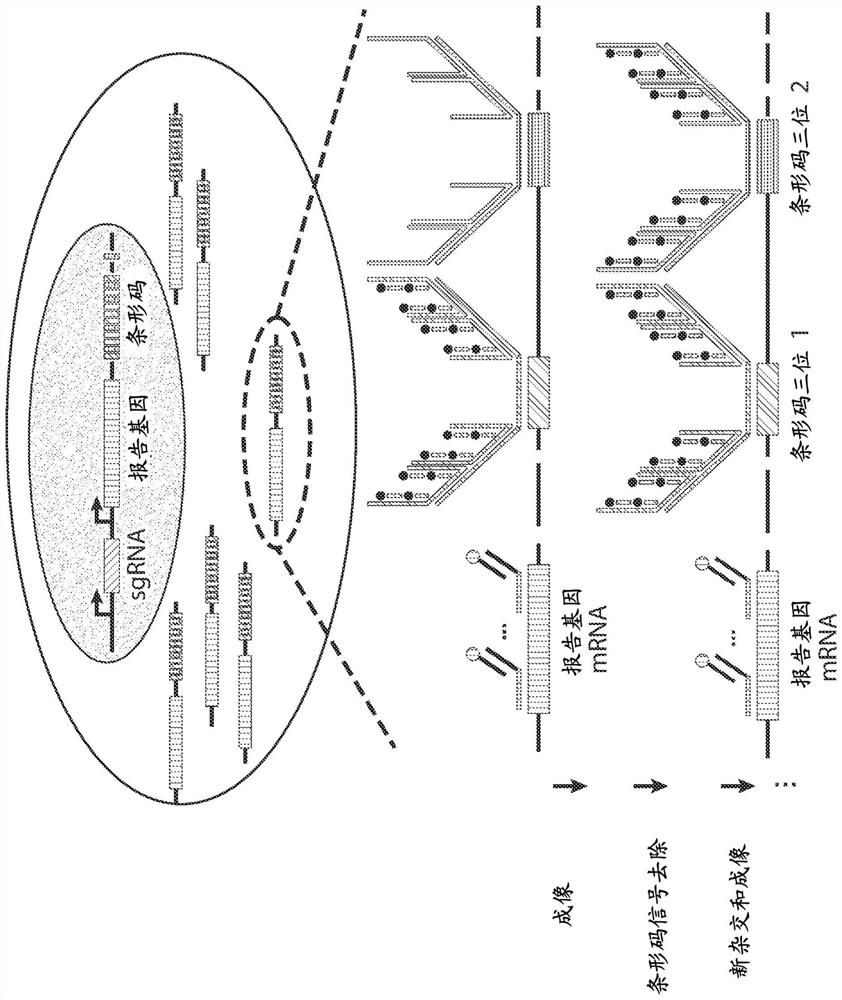
[0177]CRISPR screening of pooled libraries provides a powerful means to discover genetic factors involved in cellular processes in a high-throughput manner. However, the phenotypes available for pooled library screening are limited. Complex phenotypes, such as cellular morphology and subcellular molecular organization, as well as their dynamics, require imaging-based readouts that cannot currently be derived using pooled library CRISPR screens. These examples demonstrate a fully imaging-based hybrid library CRISPR screening approach that combines high-content phenotypic imaging with high-throughput guide RNA (sgRNA) identification in single cells. In one such approach, sgRNAs are co-delivered to cells with corresponding barcodes placed in the 3' untranslated region (3'UTR) of the reporter gene using a lentiviral delivery system with reduced recombination Induced sgRNA-barcode mismatches. Barcodes can be read out using multiplex error robust fluorescence in situ hybridization...

Embodiment 2

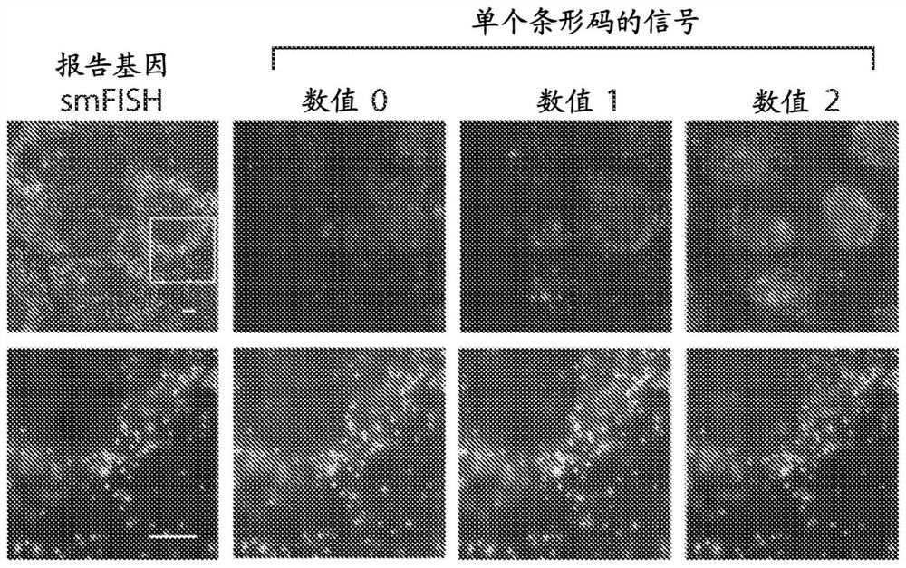
[0181] This example illustrates high-throughput high-precision barcode imaging in mammalian cells. In situ imaging-based screening of pooled libraries has been performed in bacteria in recent years, where the genotype of individual cells is identified by multiplex FISH imaging of barcodes associated with gene variants. Because of the small size of bacterial cells, the diffuse signal from barcoded RNA in a single cell is strong enough to be easily measured. However, mammalian cells are approximately thousand times larger than bacteria, making it difficult to achieve high enough concentrations of barcoded RNA to allow reliable measurements. Therefore new barcode expression and detection protocols are needed for mammalian cells to increase barcode signal and reduce background.

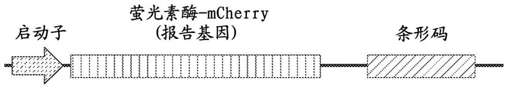
[0182] To achieve this goal, two independent promoters were used to express the sgRNA and the reporter gene in the same vector and a 12-digit ternary barcode ( Figure 1A ). Each digit (digit) of the t...

Embodiment 3

[0190] To further verify the low mislabeling rate, in this example, two reporter gene-barcode libraries were designed, each expressing a reporter with a different epitope tag (HA tag or Myc tag) fused to the barcode library as described above. gene luciferase-mCherry ( Figure 2A ), and the two libraries were cloned separately. Each library was narrowed down to contain Figure 2B ), the barcodes associated with each cell were imaged using successive rounds of hybridization as described above. The rationale is that determining the phenotype of each cell will allow the barcode identity of that cell to be deduced from the sequencing results, and then comparison with barcodes determined by imaging will allow the determination of a small fraction of misidentified barcodes.

[0191] Judging by barcode-phenotype mismatches, only ~1% of cells had incorrectly identified barcodes ( Figure 2C and 2D ). Even this small error is likely due to errors in cell segmentation, which in turn...


PUM


Abstract

The present invention generally relates to imaging cells, for example, to determine phenotypes and/or genotypes in populations of cells, e.g., to build genotype-phenotype corresponse for high-throughput screening. In some cases, the cells may be manipulated, e.g., using CRISPR or other techniques. In certain embodiments, nucleic acids may be introduced to the cell, e.g., using a lentivirus. The nucleic acids may contain a guide portion comprising a DNA or RNA recognition sequence, a reporter portion, and an identification portion comprising one or more read sequences. The guide portion may be used to alter the phenotype of the cells, e.g., using a sequence, e.g., an sgRNA sequence, that can be targeted using CRISPR or other techniques, and in some cases, the phenotype of the cells may be determined using various imaging approaches. The identification portion may be determined using MERFISH or other suitable techniques. In addition, in some cases, association or colocalization between determination of the reporter and the read sequences may substantially improve decoding accuracy, e.g., due to lowered misidentification of background signals. Other aspects are generally directed to compositions or devices for use in such methods, kits for use in such methods, or the like.

Description

[0001] related application [0002] This application claims U.S. Provisional Patent Application Serial No. 62 / 836,578, filed April 19, 2019, by Zhuang et al., entitled "Imaging-Based Pooled CRISPR Screening," and Zhuang et al., filed May 1, 2019, entitled " Imaging-Based Pooled CRISPR Screening," U.S. Provisional Patent Application Serial No. 62 / 841,715. The entire content of each of these applications is incorporated herein by reference. [0003] Government funding [0004] This invention was made with government support under Grant No. MH113094 awarded by the National Institutes of Health. The government has certain rights in this invention. technical field [0005] The present invention generally relates to imaging cells, eg, for determining the phenotype and / or genotype of a population of cells. In some cases, cells can be manipulated, eg, using CRISPR or other techniques. Background technique [0006] The development of CRISPR-based gene editing systems has greatly...

Claims


Application Information

Patent Timeline
IPC IPC(8): C12N15/113C12N9/22
CPCC12N9/22C12N2310/20C12N2320/12C12N15/113C12N15/1086C12Q1/6897C12Q1/6816C12N15/1079C12N15/1065C12Q2521/301C12Q2537/143C12Q2543/10C12Q2563/107C12Q2563/179C12N15/111G01N21/6428G01N33/52G01N2021/6439
Inventor 庄小威王翀陆天
Owner PRESIDENT & FELLOWS OF HARVARD COLLEGE
Why Eureka
Try Eureka