Multi-thread fusion living body ultrasonic thermal strain imaging method and device

An imaging method and thermal strain technology, applied in the field of medical image processing, can solve problems such as difficulty in physiological movement, poor signal-to-noise ratio, dependence on thermal strain imaging accuracy, etc., and achieve the goal of improving time resolution, enhancing reliability and robustness Effect

Pending Publication Date: 2022-04-12
NANJING UNIV
View PDF0 Cites 0 Cited by
  • Summary
  • Abstract
  • Description
  • Claims
  • Application Information

AI Technical Summary

Problems solved by technology

[0006] Aiming at the problems existing in the prior art that thermal strain imaging accuracy depends on a single motion phase, and physiological motion is difficult to be fully suppressed, resulting in thermal strain profile distortion and poor signal-to-noise ratio, while conventional spatial filtering sacrifices spatial r

Method used


Image

  • Multi-thread fusion living body ultrasonic thermal strain imaging method and device
  • Multi-thread fusion living body ultrasonic thermal strain imaging method and device
  • Multi-thread fusion living body ultrasonic thermal strain imaging method and device

Examples


Embodiment 1

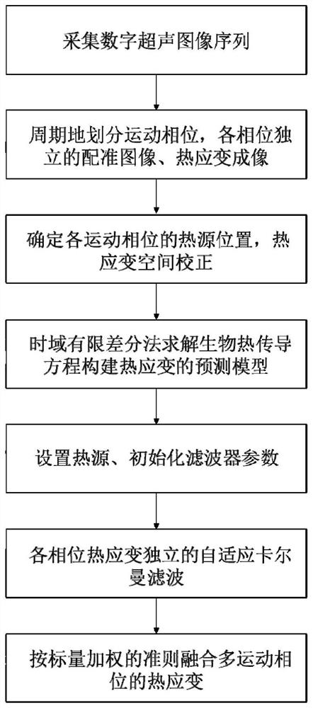
[0071] combine figure 1 As shown, the in vivo ultrasonic thermal strain imaging method of multi-thread fusion of the present invention comprises the following steps:

[0072] Acquisition steps: use hyperthermia to heat the target area, collect ultrasonic images of the living hyperthermia target area, and obtain digital ultrasonic image sequences.

[0073] Acquisition of digital ultrasound image sequences of living target tissue, specifically, the B-ultrasound instrument works in real-time imaging mode, adjust the probe angle and imaging system configuration, so that the target area is located in the imaging field of view; it is worth noting that, according to the embodiment, the body surface to Depending on the depth of the target tissue and the condition of the acoustic path, imaging probes of different types, numbers of array elements, and geometric structures can be selected, including 128 / 256 array element linear array, convex array probe, and 80 array element phased array...

Embodiment 2

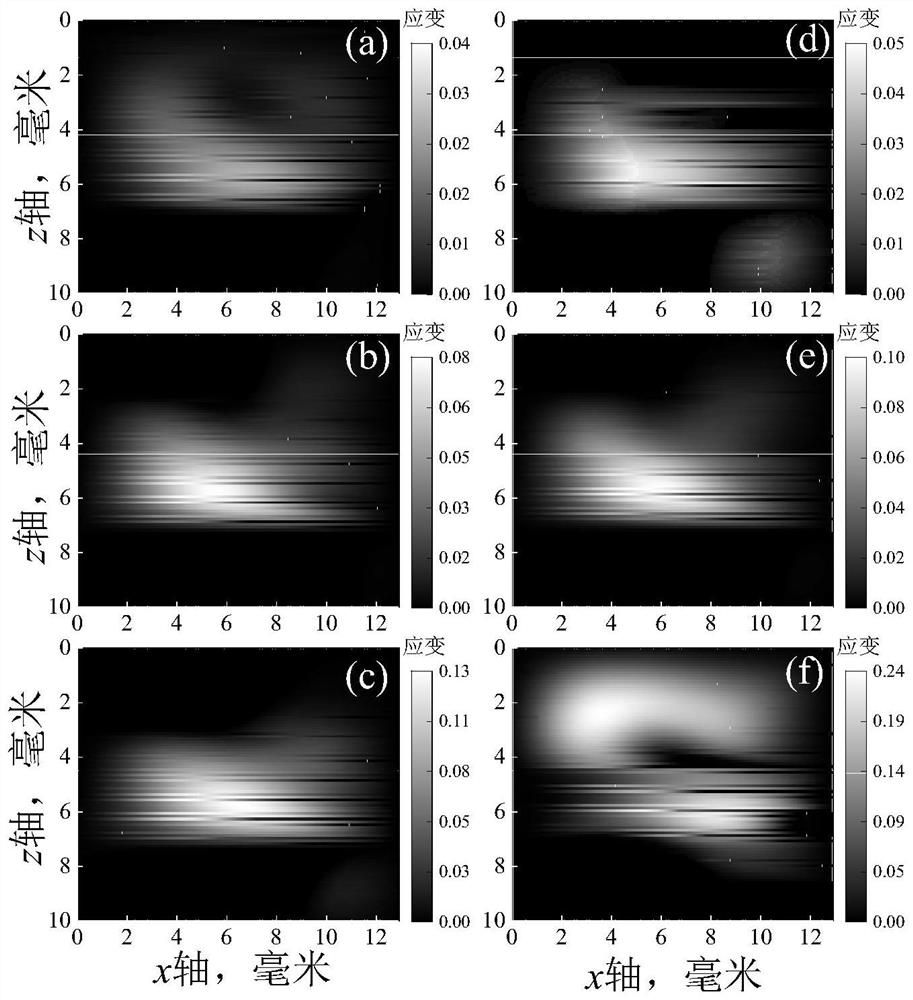
[0132] This example adopts the method in Example 1, uses microwave ablation to heat the visceral fat of living pigs, and calculates the thermal strain during the heating process. The specific steps of the method are as follows:

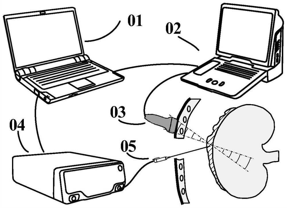
[0133] Step 1, such as figure 2 As shown, in the process of biological tissue heating and ultrasonic image acquisition, the computer 01 controls the image acquisition of the B-ultrasound imager 02 and the power emission of the microwave heater 04; the sampling rate of the B-ultrasound imager is 40MHz, with 128 array elements, center The linear array probe 03 with a frequency of 10.5MHz, the imaging depth is set to 4cm, and focused beam imaging is adopted by column-by-column scanning; the microwave ablation needle 05 is inserted into the fat tissue along the normal direction of the B-ultrasound imaging plane, and its power emission area is located on the ultrasound imaging plane; The average frame rate is 50 Hz and the ultrasonic images are collected ...


PUM

PropertyMeasurementUnit
Densityaaaaaaaaaa

Abstract

The invention discloses a multi-thread fusion living body ultrasonic thermal strain imaging method and device, and belongs to the field of medical image processing. According to the method, after an ultrasonic image sequence is collected, images are divided into a plurality of threads according to correlation, and image registration and thermal strain imaging are carried out. Furthermore, a biological heat conduction equation solved by a time domain finite difference method is used as a prediction model, and a self-adaptive Kalman filter is used for filtering the thermal strain sequence. And finally, fusing the thermal strain of each thread according to a scalar weighted linear minimum variance information fusion criterion. According to the method, the defects that in existing living body thermal strain imaging, an imaging result depends on a single motion phase, physiological motion is difficult to accurately compensate, so that thermal strain contour distortion and a poor signal-to-noise ratio are caused, and conventional spatial filtering is at the cost of sacrificing spatial resolution are overcome, and a living body thermal strain imaging result which is accurate, high in robustness and high in temporal-spatial resolution can be obtained.

Description

technical field [0001] The invention relates to the field of medical image processing, and more specifically, to a method and device for in vivo ultrasonic thermal strain imaging with multi-thread fusion. Background technique [0002] Ultrasonic thermal strain imaging is a method for quantitatively estimating the temperature by analyzing the movement of tissue scattering points in the heating process image, and then obtaining the thermally induced displacement and strain of the tissue. However, in vivo thermal strain imaging, the movement of the living body between images will lead to large strain artifacts, and even cause thermal strain imaging to fail. In order to overcome the influence of living body motion, existing thermal strain imaging methods usually use image registration or spatial filtering methods. However, due to the lack of motion phase analysis, the motion suppression effect of image registration is easily affected by the motion state of the initial image, an...

Claims


Application Information

Patent Timeline
IPC IPC(8): G06T7/00G06T7/246G06T7/277
Inventor 郭霞生尹楚豪章东屠娟
Owner NANJING UNIV
Why Eureka
Try Eureka