
Application of reagent for detecting spatial distribution of immune checkpoints in preparation of tumor treatment and prognosis products

An immune checkpoint and spatial distribution technology, applied in the field of tumor diagnosis, can solve the problems of insufficient evidence, lack of accurate prediction of the efficacy of immunotherapy in tumor patients, and unsatisfactory immunotherapy effect.

Pending Publication Date: 2022-05-17
成都福实生物科技有限公司
View PDF0 Cites 0 Cited by
  • Summary
  • Abstract
  • Description
  • Claims
  • Application Information

AI Technical Summary

Problems solved by technology

Insufficient evidence to further confirm the efficacy of immunotherapy in the TMB-High predictive phase in solid tumors, including gastrointestinal tumors
The lack of accurate prediction of the efficacy of immunotherapy in tumor patients is an important reason for the unsatisfactory effect of immunotherapy. At present, there is an urgent need to develop predictive indicators with clinical guiding value to help and guide clinicians in making decisions about the indications, timing and prognosis of immunotherapy. make an accurate judgment

Method used


Image

  • Application of reagent for detecting spatial distribution of immune checkpoints in preparation of tumor treatment and prognosis products
  • Application of reagent for detecting spatial distribution of immune checkpoints in preparation of tumor treatment and prognosis products
  • Application of reagent for detecting spatial distribution of immune checkpoints in preparation of tumor treatment and prognosis products

Examples


Embodiment 1

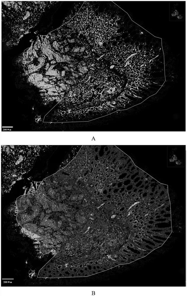
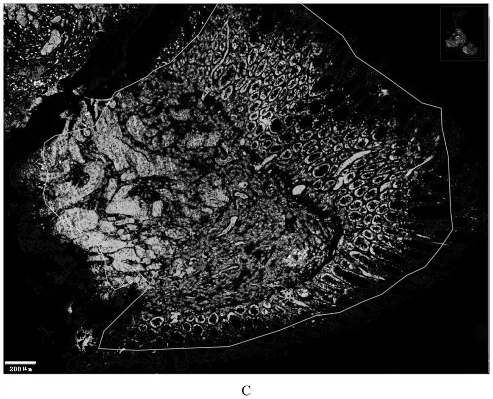
[0025] Samples including stage I-IV tumor lesions, paracancerous tissues, and tissue samples before and after immunotherapy were taken, and tissue HE staining was used to preliminarily determine the spatial characteristics of the tissue, grouped and numbered, and a follow-up cohort study was carried out at the same time.

[0026] Then multiple fluorescent staining was applied to the tumor tissue and paracancerous tissue samples for molecular markers TIM-3, PD-1, CD8, PAN-CK, DAPI staining (Opal assay kit, PerkinElmer), and Vectra Polaris (PerkinElmer) for Slice panoramic scanning imaging, the specific steps are as follows:

[0027] 1) Dewax, bake the slices in a 60°C oven for 1 hour, soak in xylene for 3 minutes and 3 times;

[0028] 2) rehydration, rehydration in gradient ethanol solutions with volume fractions of 100%, 95%, 70%, and 50% respectively;

[0029] 3) For antigen retrieval, select the antigen retrieval solution (citric acid or EDTA) according to the requirements ...

Embodiment 2

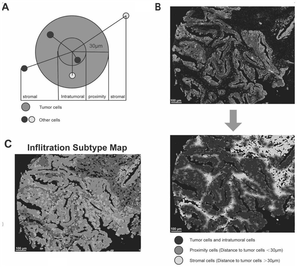
[0045] Apply artificial intelligence, machine learning and other bioinformatics technologies to explore the distribution and interaction between TIM-3 and other immune checkpoints such as PD-1 in the tumor microenvironment, as well as the distribution and interaction of extracellular matrix and immune cells in the microenvironment, and analyze tumor subtypes , to establish a spatial orientation ( figure 2 ).

[0046] In order to establish a predictive model for the spatial interaction of novel immune checkpoints ( image 3 ), first randomly divide tumor patients into two groups: training cohort and verification cohort, according to immune checkpoints and the spatial state of immune cells, we get 21 types of immune cell states, and then according to these 21 spatial states and clinical TNM staging, we establish LASSO-COX model ( image 3 , a), and defined six of the 21 spatial states (intratumoral CD8+TIM3+, intratumoral CD8+PD1+, proximity TIM3+, intratumoral PD1+, distal P...

Embodiment 3

[0048] Taking human rectal cancer patients as an example, the nomogram was used as a quantitative tool ( Figure 4 , a), through the establishment of spatial immune checkpoint TIM-3, PD-1 interaction to establish a prediction model to predict the patient's 3-year and 5-year mortality, successfully predicting the prognosis of rectal cancer patients, the results are as follows Figure 4 , b, the predicted likelihood is consistent with the actual observed likelihood.


PUM


Abstract

The invention discloses an application of a reagent for detecting spatial distribution of immune checkpoints in preparation of a tumor treatment and prognosis product, and a state-of-the-art spatial proteomics analysis technology and machine learning are adopted in the invention, so that the immune checkpoints can be used for detecting the spatial distribution of the immune checkpoints, and the immune checkpoints can be used for detecting the spatial distribution of the immune checkpoints. Spatial distribution of an immune checkpoint TIM-3 (T cell immune globulin and mucoprotein domain protein-3), an immune checkpoint PD-1 (programmed death factor-1) and the like in a solid tumor tissue tumor microenvironment is analyzed, and spatial interaction of TIM-3 molecules and immune cells / extracellular matrixes in the microenvironment is further deeply analyzed; an accurate prediction model is established by applying machine learning and the like and is applied to clinical treatment curative effect and prognosis monitoring of the generic cancer species, and the prediction model disclosed by the invention is remarkably superior to prediction of prognosis of tumor patients by traditional tumor TNM staging, and has a great application prospect in the biological field and medicine transformation research.

Description

technical field [0001] The invention relates to the field of tumor diagnosis, in particular to the application of reagents for detecting the spatial distribution of immune checkpoints in the preparation of tumor treatment and prognosis diagnosis products. Background technique [0002] Currently, the most widely studied and used immune checkpoint inhibitors include monoclonal antibodies against CTLA-4, PD-1 and PD-L1. Checkpoint immunotherapy has shown significant efficacy in some tumors, such as melanoma, but current immune checkpoint inhibitors and cell therapy have little effect on solid tumors, including pancreatic cancer. Taking pancreatic cancer as an example, in the immunotherapy of pancreatic cancer, immune checkpoint inhibitors (PD-1, PD-L1, CTLA-4) have carried out the most clinical trials. Pancreatic cancer patients have little effect. [0003] The current standard for immunotherapy detection is to analyze the expression of PD-L1 protein by immunohistochemistry. ...

Claims


Application Information

Patent Timeline
Patent Type & Authority Applications(China)
IPC IPC(8): G01N33/574G01N33/569G01N33/577G01N33/58G16B25/10G06F30/27
CPCG01N33/57484G01N33/57492G01N33/56966G01N33/577G01N33/582G16B25/10G06F30/27G01N2333/70503G01N2333/70521G01N2333/70517G01N2333/4742G06F2119/02
Inventor 姜红王平孔昊谢带黄子芮吴淳炜
Owner 成都福实生物科技有限公司
Learn More