MDA-MB-231 cell exosome detection method based on two-color co-localization and application

A technology of MDA-MB-231 and detection method, which is applied in the field of immunoassay and fluorescence imaging, can solve problems such as clinical application difficulties, and achieve good accuracy, high sensitivity and robustness

Pending Publication Date: 2022-07-05
SOUTHEAST UNIV
View PDF0 Cites 0 Cited by
  • Summary
  • Abstract
  • Description
  • Claims
  • Application Information

AI Technical Summary

Problems solved by technology

However, due to the temporal and spatial heterogeneity of its expression, and its detection req

Method used


Image

  • MDA-MB-231 cell exosome detection method based on two-color co-localization and application
  • MDA-MB-231 cell exosome detection method based on two-color co-localization and application

Examples


Embodiment

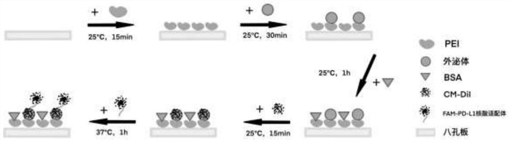
[0037] Take an eight-well plate and add 200 μL of PEI diluted 100 times to the eight-well plate. PEI is a positively charged polymer. After 15 minutes of reaction, wash with PBS three times to wash away excess PEI. Afterwards, an appropriately diluted exosome solution or a PBS solution for blank control was added. The exosome solution used was negatively charged, reacted at room temperature for 30 minutes, and rinsed with PBS. The exosome samples immobilized on the eight-well plate by electrostatic adsorption can be obtained.

[0038] The above-treated eight-well plate was subjected to excess charge blocking treatment, and 1% BSA was added to block for 1 hour at room temperature to reduce the non-specific adsorption of probes and exosomes, and PBS was rinsed. Then, 10 μg / mL of CM-DiI was dissolved in 400 μL of PBS, dropped onto an eight-well plate, reacted for 15 minutes, and the excess membrane dye was washed away by PBS. Membrane-stained exosome samples can be obtained.

...


PUM


Abstract

The invention discloses an MDA-MB-231 exosome detection method based on two-color co-localization and application. The detection method comprises the following steps: marking an MDA-MB-231 cell exosome through a cell membrane probe CM-DiI; meanwhile, a PD-L1 nucleic acid aptamer probe with another fluorophore FAM is used, and the probe can specifically capture exosomes secreted by MDA-MB-231 cells highly expressed by PD-L1; the captured exosome can be subjected to fluorescence imaging through a total internal reflection (TIRF) imaging technology; and by combining a two-color fluorescence co-localization technology, a false positive event can be judged on a single particle level, and the accuracy of an exosome fluorescence immunoassay technology is improved.

Description

technical field [0001] The invention relates to the fields of immunodetection and fluorescence imaging, in particular to an exosome detection method and application based on two-color co-localization. Background technique [0002] Regarding the detection methods of cancer, at present, commonly used clinical imaging techniques include MRI, ultrasound, CT, biopsy, etc. However, there are disadvantages such as high inspection costs and radiation harmful to the body. At the same time, when there are no obvious symptoms in the early stage, patients generally do not choose to do these inspections, which leads to the lack of early cancer treatment to some extent. Compared with ultrasound, MRI, and CT, fluorescence imaging has high sensitivity and good selectivity. The substance transitions from the ground state to the excited state after absorbing the excitation light of a certain wavelength, and then emits light with a longer wavelength than the excitation light when it returns t...

Claims


Application Information

Patent Timeline
IPC IPC(8): G01N21/64G01N33/58G01N33/574G01N33/533
CPCG01N21/6428G01N21/6458G01N33/582G01N33/574G01N33/533G01N2021/6439
Inventor 宗慎飞黄乐涵孙腾霄宗启航石晓琦
Owner SOUTHEAST UNIV
Why Eureka
Try Eureka