Methods instruments and materials for chondrocyte cell transplantation

一种软骨细胞、软骨的技术,应用在软骨细胞移植领域,能够解决缺乏功能性组织修复生物力学特性等问题

Inactive Publication Date: 2001-11-21
VERIGAN TRANSPLANT SERVICES INT (VTSI) AG
View PDF0 Cites 4 Cited by
  • Summary
  • Abstract
  • Description
  • Claims
  • Application Information

AI Technical Summary

Problems solved by technology

In all instances, the main problem was the lack of biomechanical properties required for functional tissue repair

Method used


Image

  • Methods instruments and materials for chondrocyte cell transplantation
  • Methods instruments and materials for chondrocyte cell transplantation
  • Methods instruments and materials for chondrocyte cell transplantation

Examples


Embodiment 1

[0068] in CO 2 Chondrocytes were cultured in the above growth medium for three weeks at 37°C in an incubator and operated in a class 100 laboratory at Verigen Transplantation Service ApS, Copenhagen, Denmark or Lübeck University, Lübeck, Germany. [Note: Compositions of other growth media can also be used to culture chondrocytes. ] Cells were trypsinized with EDTA for 5 to 10 minutes and counted by trypan blue viability staining in a Bürker-Türk chamber. Adjust the cell number to 7.5 x 10 per mL 5 cartilage cells. Place a piece of NUNCLON in said level 100 lab TM Plate open the cover.

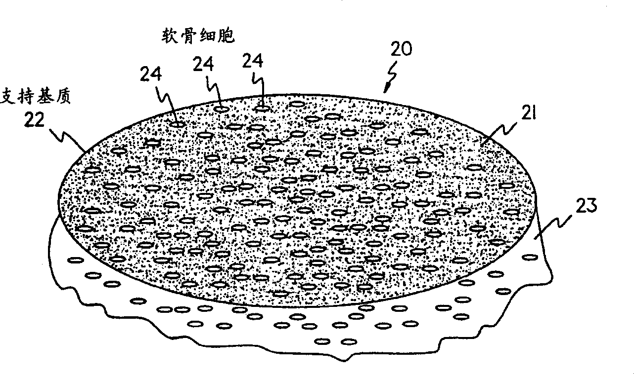
[0069] A support matrix material, specifically Chondro-Gide  Collagen membrane cut into with NUNCLON TM An appropriate size that fits the bottom of a well in a cell culture dish. In this example, a circular membrane approximately 4 cm in size was placed at the bottom of the well under aseptic conditions.

[0070] Three weeks later, the chondrocytes were transferred from the growth mediu...

Embodiment 2

[0073] in CO 2 Chondrocytes were cultured in the above growth medium for three weeks at 37°C in an incubator and operated in a class 100 laboratory at Verigen Transplantation Service ApS, Copenhagen, Denmark or at the University of Lübeck, Germany. Cells were trypsinized with EDTA for 5 to 10 minutes and counted in a Bürker-Türk chamber stained with trypan blue viability. Adjust the cell number to 5 x 10 per mL 5 cartilage cells. Place a piece of NUNCLON in the level 100 lab TM Plate open the cover.

[0074] As in Example 1, the Chondro-Gide  Support matrix cutting into and NUNCLON TM An appropriate size that fits the bottom of a well in a cell culture dish. In this example, a circular membrane approximately 4 cm in size was placed at the bottom of the well under aseptic conditions.

[0075] Three weeks later, the chondrocytes were transferred from the growth medium to the graft medium described above, and approximately 5 × 10 5 Place the individual chondrocytes direc...

Embodiment 3

[0079] in CO2 Chondrocytes were cultured in the above growth medium for three weeks at 37°C in an incubator and operated in a class 100 laboratory at Verigen Transplantation Service ApS, Copenhagen, Denmark or at the University of Lübeck, Germany. Chondrocytes were trypsinized with EDTA for 5 to 10 minutes and counted in a Bürker-Türk chamber with a trypan blue viability stain. Adjust the cell number to 5 x 10 per mL 5 cartilage cells. Place a piece of NUNCLON in the level 100 lab TM Plate open the cover.

[0080] As in Example 1, the Chondro-Gide  Support matrix cutting into and NUNCLON TM An appropriate size that fits the bottom of a well in a cell culture dish. In this example, a circular membrane approximately 4 cm in size was placed at the bottom of the well under aseptic conditions.

[0081] Three weeks later, the chondrocytes were transferred from the growth medium to the graft medium described above, and approximately 5 × 10 5 Place the individual chondrocytes ...


PUM


Abstract

The present invention provides a method for the effective treatment of articulating joint surface cartilage in an animal by the transplantation of an implantable article including chondrocyte cells retained to an absorbable support matrix. The present invention is also directed to an instrument for placing and manipulating the implantable article at the site of implantation, and a retention device for securing the implantable article to the site of implantation. The present invention is also directed to an implantable article for cartilage repair in an animal, the implantable article including chondrocyte cells retained on an absorbable support matrix, and a method of making same. This invention also encompasses, in general, an article comprising an absorbable flexible support matrix for living cells grown and adhered thereto.

Description

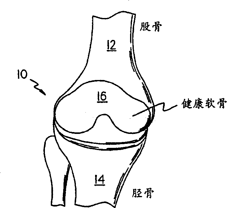
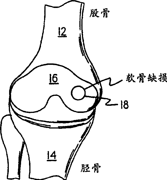
field of invention [0001] The present invention relates to chondrocyte transplantation, bone and cartilage transplantation, healing, joint repair and prevention of arthritic conditions. In particular, the present invention relates to new methods and tools for chondrocyte transplantation and cartilage regeneration. Background of the invention [0002] More than half a million arthroplasties and total joint replacements are performed in the United States each year. About the same number of procedures are performed in Europe. The European caseload includes around 90,000 total knee replacements and 50,000 surgeries to repair knee defects. These case numbers are essentially the same in the United States (Praemer A., ​​Fumer S., Rice, D.P., State of the American Musculoskeletal, American Academy of Orthopedic Surgeons, Park Ridge, Ill., 1992, 125). [0003] Approaches for cartilage regeneration therapy would be most effective and be performed at an early stage of joint damage, ...

Claims


Application Information

Patent Timeline
Patent Type & Authority Applications(China)
IPC IPC(8): A61B17/56A61B17/00A61B17/06A61B17/064A61B17/22A61B17/68A61B17/86A61F2/00A61F2/02A61F2/28A61F2/30A61F2/38A61F2/46A61K9/00A61K35/32A61K47/42A61L24/00A61L27/38A61L31/04
CPCA61F2002/30028A61L2430/06A61F2002/30062A61B17/06166A61F2002/30761A61F2002/009A61F2002/4627A61B17/0642A61F2002/30764A61F2/30756A61L31/04A61L27/3817A61F2310/00365A61F2002/30013A61B17/00491A61F2002/30006A61B17/68A61F2230/0091A61F2002/4624A61F2/38A61F2220/005A61F2002/30932A61F2/3094A61F2002/30762A61B17/86A61B2017/00004A61F2250/0051A61F2250/0015A61F2250/0024A61F2/4618A61F2002/30293A61B17/221A61F2002/4635A61B2017/2215A61F2002/30448A61L27/3852A61F2210/0004A61B2017/0647C08L67/04A61F2002/30011A61F2/4603A61L27/58A61L27/22A61L27/24
Inventor 布鲁诺·贾内蒂彼得·贝伦斯塞缪尔·阿斯库莱
Owner VERIGAN TRANSPLANT SERVICES INT (VTSI) AG
Why Eureka
Try Eureka