

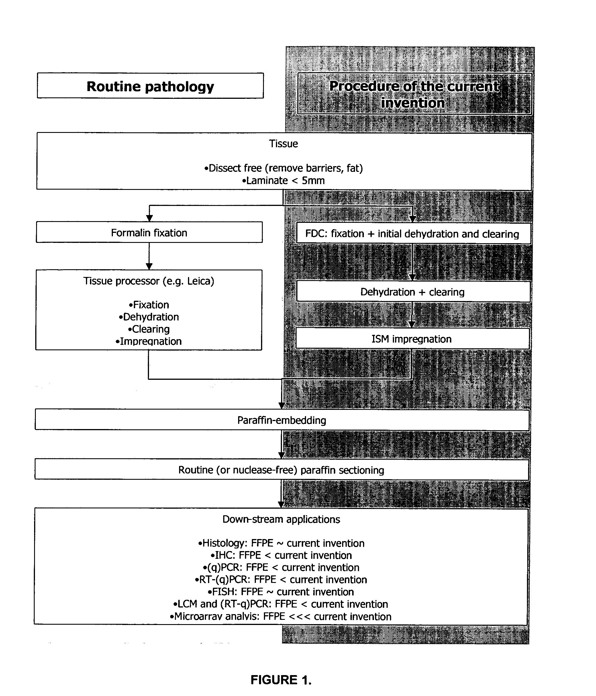
Methods, Reagents, Devices and Instrumentation For Preparing Impregnated Tissue Samples Suitable For Histopathological and Molecular Studies
a tissue sample and histopathological technology, applied in the field of histopathological and molecular studies, can solve the problems of affecting the morphology of frozen tissue samples, affecting the morphology of paraffin-embedded tissue samples, and severely compromising gene expression data, and achieve the effect of low melting point paraffin
- Summary
- Abstract
- Description
- Claims
- Application Information
AI Technical Summary
Benefits of technology
Problems solved by technology
Method used
Image

Examples
example 1
Exemplary General Manner of Tissue Processing
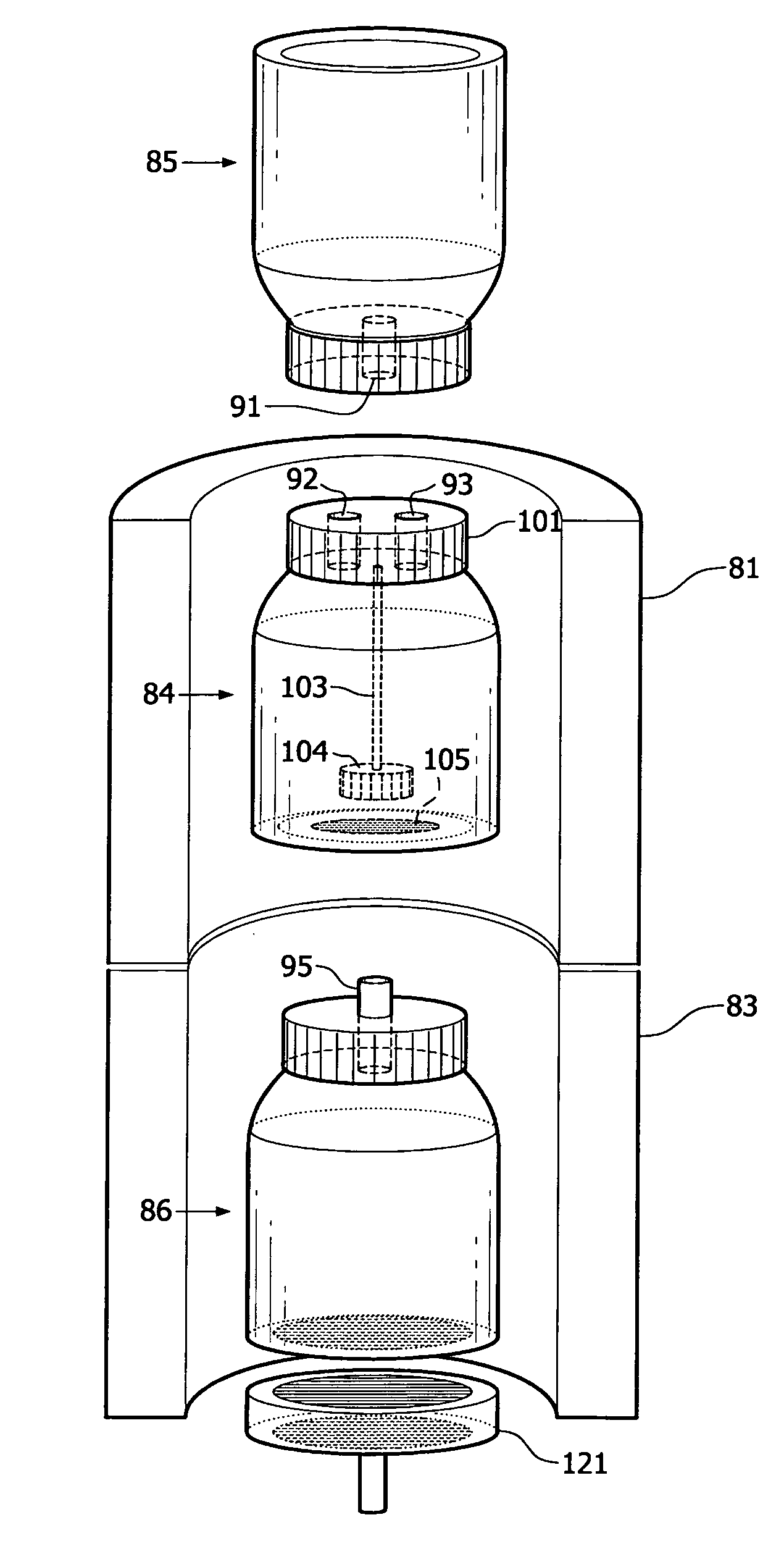
[0267]Tissue samples to be processed according to the current invention should be processed as quickly as possible after excision from the source after the onset of ischemia or death, or after removal from the soil or any other substrate solution or medium. In the case the samples posses a protective barrier that would interfere with FDC diffusion (e.g. a renal capsule or the waxy coating of plant material) this barrier should be removed prior to proceeding with the processing as described in the current invention. Large samples should be dissected / laminated into smaller fragments to maximize FDC diffusion in the first step of the present method. A general rule of thumb states the tissue thickness should not exceed 5 mm in at least one spatial dimension to allow proper fixation (Kieman J P: Histopathological and Histochemical Methods, Theory, and Practice, ed 3. Oxford, Butterworth-Heinemann, 1999). Samples that consist of cell cultures s...
example 2
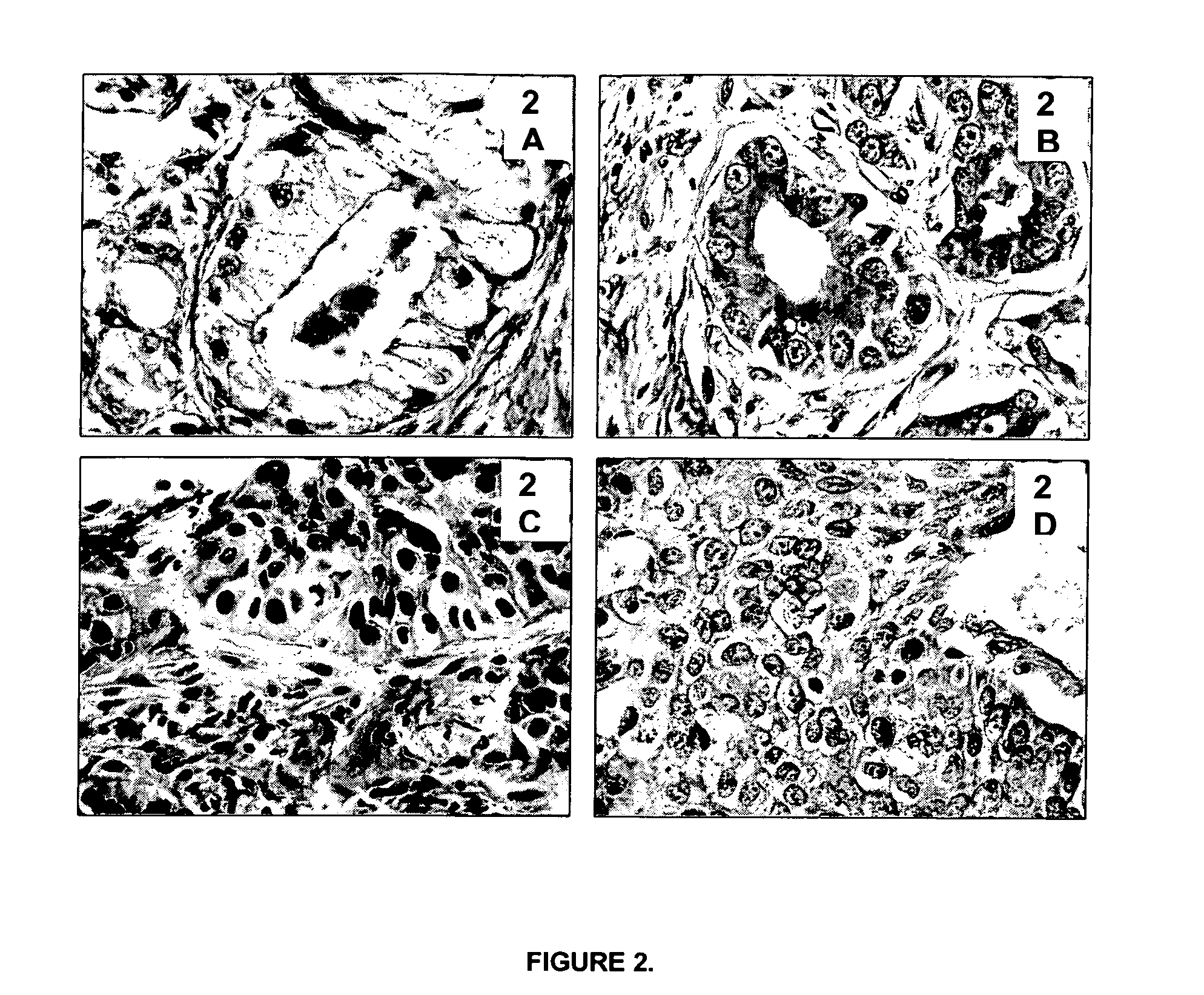
The Tissue Morphological Detail and Cytology of Tissues Processed According to the Current Invention is as Good as or Better than FFPE Tissues (e.g. Prostate Tissue and Bronchoscopic Biopsy)
[0268]A prerequisite for the general acceptance and introduction of the current invention into routine pathology, research and clinical laboratories is that tissues processed according to the described invention have a morphology comparable or superior to the one of FFPE tissues.
[0269]Typical examples that demonstrate the importance of preservation of good morphological detail for histopathological diagnosis are prostatic and bronchoscopic biopsies. Prostatic (FIGS. 2A and 2B) and bronchoscopic biopsies (FIGS. 2C and 2D) are shown. FIGS. 2A and 2C show biopsies processed according to standard routine pathology procedures (4% formalin fixation for 16 hours at room temperature, followed by tissue processing in a Leica Tissue Processor). FIGS. 2B and 2D show biopsies processed according to the proce...
example 3
Immunohistochemical (IHC) Stainings Performed on Tissue Processed According to the Described Invention and Embedded in Paraffin
[0271]The antigenic profile of the samples processed according to the current invention should be comparable to the one of FFPE tissues. In addition, the antigen retrieval procedures that are applied to FFPE tissues should be applicable without any (or only minor) adaptations on material processed according to the described invention. Table 1 lists 85 antibodies (both monoclonal and polyclonal) that have been applied successfully on human surplus tissues processed according to the described invention. In total, 4239 sections were stained immunohistochemically and evaluated by pathologists, who found no differences between standard FFPE sections and sections from tissues processed according to the current invention. Data is shown in Table 1.
TABLE 1Number ofIHC stainingsType ofAntibody targetperformedantibody-1-antitrypsin10P-actin, sarcomeric6M-smooth muscle ...
PUM
 Abstract
Description
Claims
Application Information