Method for preparation of neo-cartilage constructs

a technology of neocartilage and constructs, which is applied in the field of neocartilage constructs, can solve the problems of debilitating disability, affecting mobility, and damage to the articular cartilage of active individuals and older generation adults

Inactive Publication Date: 2006-09-28
TAKAGI IND CO LTD +1
View PDF24 Cites 29 Cited by
  • Summary
  • Abstract
  • Description
  • Claims
  • Application Information

AI Technical Summary

Benefits of technology

[0036] d) depositing a space-holding thermo-reversible gel (SHTG) or TRGH into a lesion or into a cavity formed above the first sealant layer thereby permitting sufficient time for growth and differentiation of ex vivo cultured neo-cartilage, said space holding thermo-reversible gel (SHTG) deposited into said cavity as a sol at temperatures between about 5 to about 25° C., wherein within said cavity and at the body temperature said SHTG converts from the fluidic sol into a solid gel and in this form SHTG holds the space for subsequent introduction of the neo-cartilage cultured ex vivo, and provides protection against cell and blood-borne agents migration into the cavity from the subchondral space and from the synovial capsule and wherein its presence further provides a substrate for and promotes in situ formation of a de novo superficial cartilage layer covering the cartilage lesion;
[0052] Still another aspect of the current invention is a method for generation and maintaining integrity of the lesion cavity for the introduction of neo-cartilage, a neo-cartilage gel, a neo-cartilage suspension or neo-cartilage construct from a synovial capsule and for blocking the migration of subchondral and synovial cells and cell and blood products into said cavity and for providing a substrate for a formation of superficial cartilage layer overgrowing the lesion by introducing a biologically acceptable space-holding thermo-reversible gel (SHTG) into a cleaned lesion for a duration of culturing autologous chondrocytes into neo-cartilage before introducing said neo-cartilage or neo-cartilage construct or suspension into the lesion.

Problems solved by technology

Damage to the articular cartilage which occurs in active individuals and older generation adults as a result of either acute or repetitive traumatic injury or aging is quite common.
Such damaged cartilage leads to pain, affects mobility and results in debilitating disability.
For example, severe and chronic forms of knee joint cartilage damage can lead to greater deterioration of the joint cartilage and may eventually lead to a total knee joint replacement.

Method used


Image

  • Method for preparation of neo-cartilage constructs
  • Method for preparation of neo-cartilage constructs
  • Method for preparation of neo-cartilage constructs

Examples


example 1

Isolation of Chondrocytes from Source Tissue

[0361] This example describes the procedure used for isolation of chondrocytes from swine cartilage.

[0362] Chondrocytes were enzymatically isolated from cartilage harvested under sterile conditions from the hind limbs of 6-month old swine. The femur was detached from the tibia and the trachea head exposed. Strips of cartilage were removed from the trachea using a surgical blade.

[0363] The cartilage was minced, digested in a 0.15% collagenase type I solution in DMEM / Nutrient Mixture F-12 (DMEM / F-12) 1:1 mixture with 1% penicillin-streptomycin (P / S) and gently rotated for 18 hours at 37° C. Chondrocytes were collected and rinsed twice by centrifugation at 1500 rpm for 5 min. Chondrocytes were re-suspended in DMEM / F-12 containing 1% penicillin-streptomysin and 10% FBS.

[0364] Chondrocytes were expanded for about 5 days at 370C.

example 2

The Production of Human Neo-Cartilage Construct

[0365] This example describes conditions for production of neo-cartilage for human use.

[0366] The patient undergoes arthroscopic biopsy of a small (200-500 mg) piece of healthy cartilage from the ipsilateral knee. The biopsy is taken from the non-weight bearing portion of the femoral condyle or from the femoral notch as deemed most appropriate for the patient. The biopsy sample is placed into a sterile, non-cytotoxic, non-pyrogenic specimen container which is packaged and shipped to the laboratory.

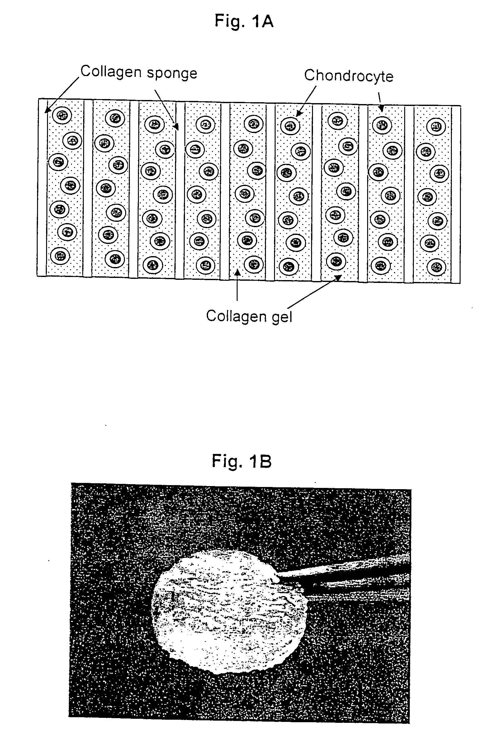
[0367] At the laboratory the biopsy sample is examined against acceptance criteria and then transferred to the chondrocyte isolation and expansion area. Samples from the biopsy specimen transport buffer are tested for sterility and for mycoplasma. The expanded chondrocytes are suspended in VITROGEN® gellable collagen solution, commercially available from Cohesion Corp., Palo Alto, Calif. A pre-formed collagen sponge (22×22 mm square and 2-4...

example 3

Preparation of Support Matrices

[0370] This example illustrates preparation of the cellular support matrix, also called the TESS matrix.

[0371] 300 grams of a 1% aqueous atelocollagen solution (VITROGEN®), maintained at pH 3.0, is poured into a 10×20 cm tray. This tray is then placed in a 5 liter container. A 50 ml open container containing 30 ml of a 3% aqueous ammonia solution is then placed next to the tray, in the 5 liter chamber, containing 300 grams of said 1% aqueous solution of atelocollagen. The 5 liter container containing the open trays of atelocollagen and ammonia is then sealed and left to stand at room temperature for 12 hours. During this period the ammonia gas, released from the open container of aqueous ammonia and confined within the sealed 5 liter container, is reacted with the aqueous atelocollagen resulting in gelling said aqueous solution of atelocollagen.

[0372] The collagenous gel is then washed with water overnight and, subsequently, freeze-dried to yield a ...


PUM

PropertyMeasurementUnit
pressureaaaaaaaaaa
pressureaaaaaaaaaa
temperaturesaaaaaaaaaa

Abstract

Neo-cartilage constructs suitable for implantation into a joint cartilage lesion in situ and a method for repair and restoration of function of injured, traumatized, aged or diseased cartilage. The construct comprises at least chondrocytes incorporated into a support matrix processed according to the algorithm comprising variable hydrostatic or atmospheric pressure or non-pressure conditions, variable rate of perfusion, variable medium composition, variable temperature, variable cell density and variable time to which the chondrocytes are subjected.

Description

[0001] This application is a continuation-in-part application of Ser. No. 10 / 104,677 filed on Mar. 22, 2002 and is based on and claims priority of the Provisional application Ser. No. 60 / 425,696 filed on Nov. 12, 2002 and Provisional application Ser. No. 60 / 427,627 filed on Nov. 18, 2002.BACKGROUND OF THE INVENTION Field of Invention [0002] The current invention concerns neo-cartilage constructs suitable for implantation into a joint cartilage lesion in situ and a method for repair and restoration of function of injured, traumatized, aged or diseased cartilage. The construct of the invention comprises at least chondrocytes incorporated into a support matrix processed according to the algorithm of the invention. In particular, the invention concerns a neo-cartilage construct for in situ implanting into a cartilage lesion wherein said construct comprises a cultured differentiated autologous or heterologous chondrocytes or cells which could be differentiated into chondrocytes incorpora...

Claims


Application Information

Patent Timeline
Patent Type & Authority Applications(United States)
IPC IPC(8): C12N5/02C12N5/00A61F2/00A61K35/12A61L27/24A61L27/38A61L27/56C12N5/077
CPCA61F2310/00365A61L2430/06A61L27/24A61L27/3633A61L27/3817A61L27/3843A61L27/3852A61L27/3895A61L27/56C12N5/0655C12N2533/54A61K35/12A61K35/32A61K9/0024A61L27/38A61F2/28A61L27/54A61L2300/64C12N2521/00
Inventor MIZUNO, SHUICHIKUSANAGI, AKIHIKOTARRANT, LAURENCE J. B.TOKUNO, TOSHIMASASMITH, ROBERT LANE
Owner TAKAGI IND CO LTD
Why Eureka
Try Eureka