

Microarray chip used for cell or microorganism detection and its preparation method
A microarray chip and microorganism detection technology, applied in biochemical equipment and methods, microorganism measurement/inspection, biological testing, etc., to achieve the effect of reducing detection costs
- Summary
- Abstract
- Description
- Claims
- Application Information
AI Technical Summary
Problems solved by technology
Method used
Image


Examples
Embodiment 1
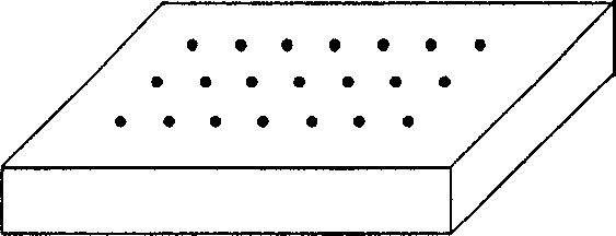
[0024] The protein microarray for detecting cells and microorganisms in this example uses polystyrene plastic plate as the base material, and the upper surface of the plate is modified with aldehyde groups, and the upper surface of the aldehyde groups has microarrays formed by various proteins.
[0025] The protein microarray-based cell detection chip of this embodiment is prepared by the following steps:
[0026] A. Put the polystyrene plastic plate in 1%-10% glutaraldehyde PBS solution, react at room temperature for 2 hours, take it out, wash it with pure water, and dry it with nitrogen;
[0027] B. Spot the required protein solution on the plate obtained in the above "A" step to complete the preparation of the protein microarray.
[0028] When in use, it is only necessary to apply the sample to be tested on the protein microarray, wash off the unbound sample solution, observe the detection result with an optical microscope, or collect images with a CCD for image analysis. ...
Embodiment 2
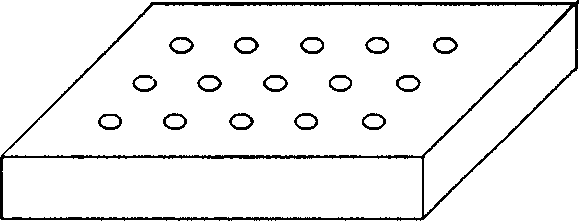
[0030] The protein microarray used to detect cells and microorganisms in this embodiment uses a gold film as the base material, and the upper surface of the gold film 1 is prepared with micro-shaped grooves, and the upper surface of the micro-shaped grooves is modified with sulfhydryl groups. There are microarrays formed by various proteins on the upper surface.
[0031] A. Clean the surface of the gold film with an acid-base cleaning solution, rinse it with pure water, and dry it with nitrogen;
[0032] B. prepare microgrooves on the gold film surface of A gained;
[0033] C. modify the sulfhydryl group on the upper surface in the microgroove;
[0034] D. Spot the desired protein solution on the glass slide obtained in C to complete the preparation of the protein microarray.
[0035] When in use, the sample solution to be detected is applied to the protein microarray, the unbound sample solution is washed, and the result is detected by an electrochemical method.
Embodiment 3
[0037] The protein microarray used to detect cells and microorganisms in this embodiment uses glass slides as the base material, and the upper surface of the glass slides is modified with agarose to form micro-projections. Sugar-formed microarrays.
[0038] The microarray for detecting cells and microorganisms of this embodiment is prepared by the following steps:
[0039] A. Place the glass slide in 95% acetone-water solution containing 1%-5% aminosilane, react at room temperature for 30 minutes, take it out, clean it with pure water, and dry it with nitrogen;
[0040] B. dissolving agarose in water, controlling the concentration at 0.5-3.0%, and coating it on the glass slide obtained in B, forming an agarose microarray C on the glass slide;
[0041] C. Cover the surface of the agarose microarray obtained in C with NaIO 4 Solution activation to generate reactive groups;
[0042] D. Immobilizing different types of proteins or oligosaccharides on the activated agarose surfac...
PUM
 Abstract
Description
Claims
Application Information