

Autoantibody detection tissue chip
A tissue chip and autoantibody technology, which is applied in the field of biomedical detection chips, can solve the problems of inability to detect autoantibodies well, the limitation of cultured cells is strong, and the tissue structure is not clear enough, and achieves a simple and feasible preparation method and is easy to commercialize. the effect of a clear organizational structure
- Summary
- Abstract
- Description
- Claims
- Application Information
AI Technical Summary
Problems solved by technology
Method used
Image

Examples
Embodiment 1
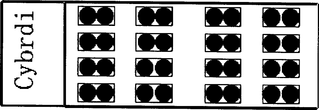
[0033] Example 1, see figure 1 ,
[0034] 1. Anti-double-stranded DNA antibody detection chip:
[0035] The matrix combination of cultured Hep-2 cells and liver tissue is selected for detection matrix. The combination of Hep-2 cells and liver tissue is the standard matrix for detecting double-stranded DNA antibodies. Two liver tissue areas are combined into one detection area, the basic unit area is 0.3cm×0.6cm, the area of Hep-2 cells is 0.3cm×0.3cm, and the number of Hep-2 cells is 5×10 2 ~1×10 3 piece / mm 2 , the size of the liver tissue area is 0.3cm×0.3cm.
[0036] There are 16 basic units arranged on the detection chip, which are arranged into a combination of 4 basic units to form a tissue / cell microarray, which can detect at least 4 different serum samples at a time or set at least 4 different serum dilutions for the same serum. detection. The liver tissue matrix is a paraffin section or a frozen section of fresh liver tissue.
[0037] 2. The production proc...
Embodiment 2
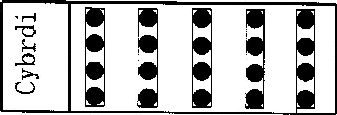
[0052] Example 2, see figure 2 : There are 5 basic units arranged on the detection chip, and each basic unit is arranged from top to bottom in order of Hep-2 cells, monkey liver, rat liver, and human embryonic liver paraffin section combination, and the size of each point is 0.3cm. The distance between each point in the vertical direction is 0.3cm, the distance between each point in the horizontal direction is 0.8cm, and the number of Hep-2 cells is 5×10 2 ~1×10 3 piece / mm 2 , 1 to 3 serums can be detected at a time.
[0053] (2) The basic unit arranged on the organ / tissue-specific antibody detection chip is the same or different single tissue or tissue combination for detection of various autoimmune antibodies. The appearance of each of the above autoantibodies is related to one or more related to an autoimmune disease, or a specific / marker antibody for a certain type of autoimmune disease, this group of chips can be used for clinical assistance in the diagnosis of autoim...
Embodiment 3
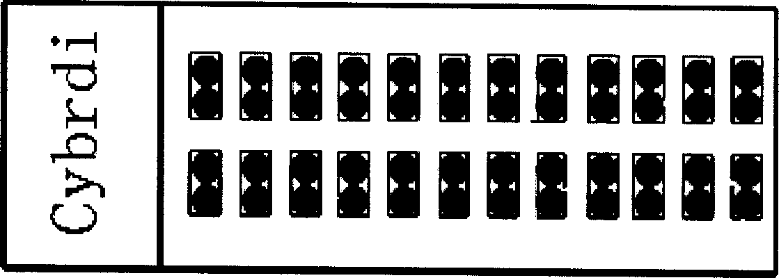
[0058] Example 3, see image 3 :
[0059] 1. Production process of anti-desmosome antibody / pemphigus antibody detection chip
[0060] The detection matrix is a combination of human (or rat) esophagus and tongue tissue paraffin sections, which is the standard matrix for detecting double-stranded DNA antibodies. Area (basic unit), the size of the basic unit is 0.3cm×0.6cm, the size of the esophageal tissue area is 0.3cm×0.3cm, and the size of the tongue tissue area is 0.3cm×0.3cm.
[0061] The arrangement of paraffin tissue slices in the detection chip is 4×12 points, each point has a diameter of 0.3 cm, and there are four points in the longitudinal direction, which are esophagus tissue, tongue tissue, esophagus tissue and tongue tissue in sequence, points 1 and 2 They are connected to form a basic unit. The 3rd and 4th points are connected to form a basic unit. The distance between the 2nd and 3rd points is 0.6cm, and there are 12 columns in the horizontal direction. Morph...
PUM
| Property | Measurement | Unit |
|---|---|---|
| pore size | aaaaa | aaaaa |
| pore size | aaaaa | aaaaa |
Abstract
Description
Claims
Application Information