

Large capacity tissue array making method
A tissue array and manufacturing method technology, which is applied in the fields of biochemical equipment and methods, material inspection products, and microorganism determination/inspection, etc., can solve the problems of insufficient expansion, high price, difficult separation of adhesive tapes, etc., and it is easy to achieve the effect of exhibition. Control, avoid RNA degradation, save reagents and time
- Summary
- Abstract
- Description
- Claims
- Application Information
AI Technical Summary
Problems solved by technology
Method used
Image

Examples
Embodiment 1
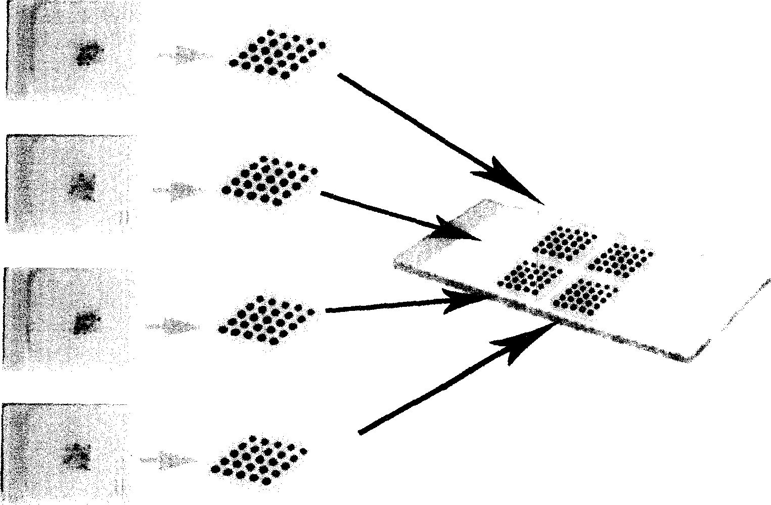
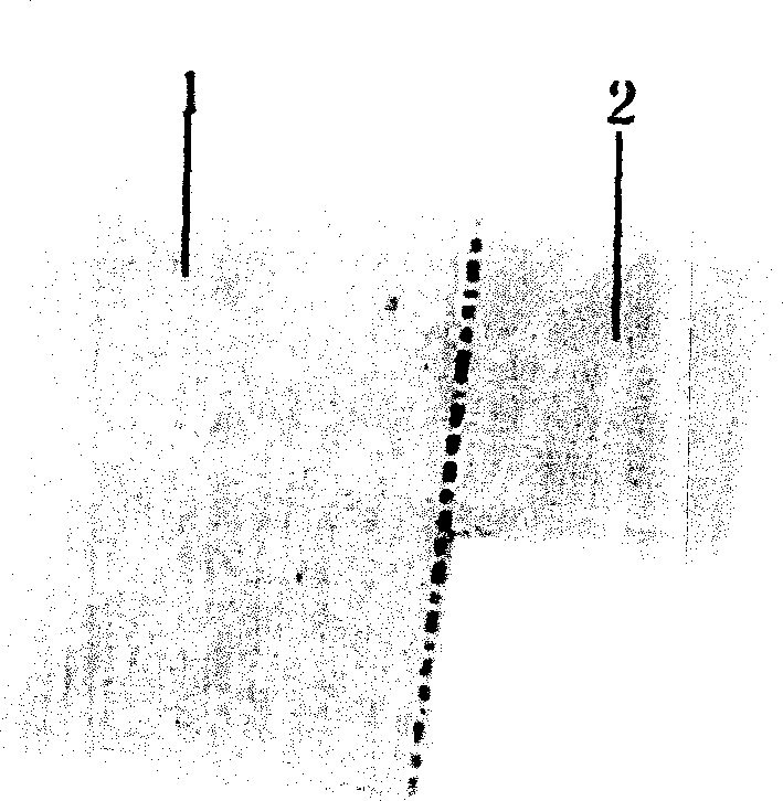
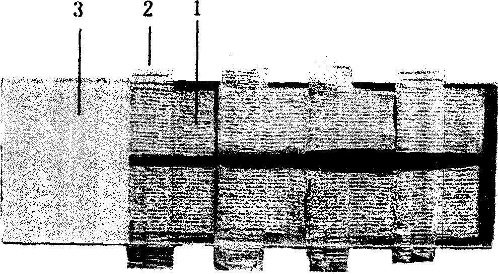
[0027] 1. Preparation of tissue array slices: 4 tissue cores were randomly selected in the marked area for each specimen. When the tissue samples are filled into the wax holes, the specific position of each sample in the two-dimensional array is recorded, and the orientation of the tissue microarray is marked at a certain position. Each blank wax block is made into 19×17 dots, and three less dots are marked in the last row as the array orientation mark. There can be 320 tissue cores in each array, and a total of 8 wax blocks containing 320 tissue cores were made. Dry-bake the 8 wax blocks made by the above method at 40°C for 10 minutes, then freeze at -20°C for 30 minutes, use a tissue slicer to produce a paraffin film containing the tissue core, and then cut off the blank wax film on the top, bottom and left of the tissue core 1 , and finally cut off the lower half of the blank wax film on the right, and keep the upper blank wax film to form a bulge 2 (see figure 2 ). The...
Embodiment 2
[0033] 1. Preparation of tissue array slices: 160 paraffin-embedded specimens of B-cell lymphoma were collected from the Department of Pathology of Nanfang Hospital from 1998 to 2004. Each specimen was sliced and stained with HE to determine the tumor concentration in the specimen. , and marked, and then 5 tissue cores were randomly selected in the marked area and filled into the acceptor wax block. Each blank wax block is made into 15×14 points, and ten points less are marked in the last row as the array orientation. There can be 200 tissue cores in each array, and a total of 4 tissue cores containing 200 tissue cores are made. of wax blocks. Dry-bake the above paraffin block at 40°C for 10 minutes, then freeze it at -20°C for 30 minutes, use a tissue slicer to make a paraffin film section containing the tissue core, and then cut off the blank wax film on the top, bottom and left side of the tissue core 1, and finally cut off The blank wax film on the lower right side, kee...
Embodiment 3
[0038] 1. Preparation of tissue array slices: 150 paraffin-embedded samples of colorectal cancer, 150 cases of paracancerous tissues, and 120 cases of metastatic lymph nodes were collected from the pathology department of Nanfang Hospital from 1998 to 2004. Each sample was sliced first, HE Stain, determine the sampling site, and mark it, then randomly select 3 tissue cores in the marked area and fill them into the acceptor wax block, and record the specific position of each sample in the two-dimensional array. Each blank wax block is made into 18×18 points, and 9 points are marked in the last row as the array orientation, so there can be 315 tissue cores in each array, and a total of 4 tissue cores containing 315 tissues were made. Wax block for wick. Dry-bake the above-mentioned paraffin block containing the tissue core in an oven at 40°C for 10 minutes, then freeze at -20°C for 30 minutes, use a tissue slicer to make a paraffin film section containing the tissue core, and ...
PUM
 Abstract
Description
Claims
Application Information