System for generating composite images for endoscopic surgery of moving and deformable anatomy

a technology of moving and deformation anatomy and composite images, which is applied in the field of minimally invasive surgeries, can solve the problems of limited surgical training, limited surgical training, and inability to give surgeons a good appreciation of depth and 3d spatial relationships among anatomical structures, and achieves enhanced understanding, reduced procedure time, and improved surgical efficiency.

Inactive Publication Date: 2019-10-01
CHILDRENS NAT MEDICAL CENT
View PDF13 Cites 21 Cited by
  • Summary
  • Abstract
  • Description
  • Claims
  • Application Information

AI Technical Summary

Benefits of technology

[0018]Utilizing emerging camera technology, a system according to the present disclosure provides a real-time stereoscopic augmented reality (AR) system for laparoscopic surgery by merging live laparoscopic ultrasound image data with stereoscopic video. The system creates two new visual cues—(1) perception of true depth with improved understanding of 3D spatial relationships among anatomical structures, and (2) visualization of critical internal structures along with a more comprehensive visualization of the operating field.
[0021]In one or more embodiments, a composite image generation device or system according to the present disclosure uses stereoscopic endoscopes based on 3D camera technology. Stereoscopic endoscopes provide two separate video streams—one each for the observer's left eye and the right eye. When viewed on an appropriate 3D display device, the resulting images give the observer a perception of depth and an enhanced understanding of 3D spatial relationship among anatomic structures. Studies suggest that being able to see the surgical field in 3D rather than in 2D provides clear advantages in terms of greater surgical efficiency (and hence reduced procedure time) and acceleration of the learning curve [2].
[0022]In one or more embodiments, LUS image data utilized for generating composite AR streams is received from an ultrasound imager implemented within an OR table. Such a capability permits continuous hands-free volumetric scanning of the patient lying on the OR table and provides the operating surgeon an unprecedented level of detail, in real time, of the surgical field. The availability of volumetric data may also ease the interpretation problem of conventional ultrasound images and allow presenting to operating surgeons 3D rendered views and reformatted cross-sectional views different from the acquisition planes.
[0025]To demonstrate the results of an embodiment of a stereoscopic AR system according to the present disclosure, a series of stereoscopic AR interrogations were conducted on the liver, gall bladder, biliary tree, and kidneys in two swine. The preclinical studies demonstrated the feasibility of the stereoscopic AR system during in-vivo procedures. Major internal structures could be easily identified. The system exhibited unobservable latency with acceptable image-to-video registration accuracy. As exhibited by these results a stereoscopic AR system according to the present disclosure provides new capabilities with respect to introducing new visual cues and enhancing visualization of the surgical anatomy, thereby improving the precision of, and expanding the capacity of, minimally invasive laparoscopic surgeries.
[0035]9) Methods for creating a real-time image augmentation system with low latency.

Problems solved by technology

Despite this success and the increasing application of minimally invasive surgery to treat or correct various pathologic conditions, visualization of the surgical field is more challenging in endoscopic surgery than in open surgery.
Furthermore, standard endoscopic video, often monocular, does not give a surgeon a good appreciation of depth and 3D spatial relationships among anatomical structure.
These limitations could be a source of major complications, which, for laparoscopic cholecystectomy, is reported to be up to 1.74% [1].
These limitations could further be attributed to minimally invasive surgeries being a relatively small share of all surgeries.
A challenge in doing so is the dynamic nature of the surgical anatomy.
However, not all imaging modalities are appropriate for use in the OR.
For example, it is difficult to perform positron emission tomography (PET) intra-operatively; however, there are instances in which combining the PET data with endoscopic video may aid the surgeon.
In addition to the laparoscopic ultrasound limitation of only showing 2D cross-sections of the 3D, its use has another limitation in that it necessitates creating an additional port and an extra hand to operate related devices.
Although real-time, conventional endoscopic video images provide a flat representation of the 3D operative field and are incapable of visualizing internal structures located beneath organ surfaces.
Computed tomography (CT) and magnetic resonance (MR) images are difficult to fuse in real time with laparoscopic views due to the deformable nature of soft-tissue organs.
In certain embodiments, the LUS image data may be 2D, which presents challenges when generating composite streams using a combination stereoscopic video overlaid with the LUS image data.
An overlay of these fundamentally different image formats makes the resulting AR difficult to interpret for laparoscopic surgeons.
The system exhibited unobservable latency with acceptable image-to-video registration accuracy.

Method used


Image

  • System for generating composite images for endoscopic surgery of moving and deformable anatomy
  • System for generating composite images for endoscopic surgery of moving and deformable anatomy
  • System for generating composite images for endoscopic surgery of moving and deformable anatomy

Examples


Embodiment Construction

[0059]Referring now to the drawings, wherein like reference numerals designate identical or corresponding parts throughout the several views.

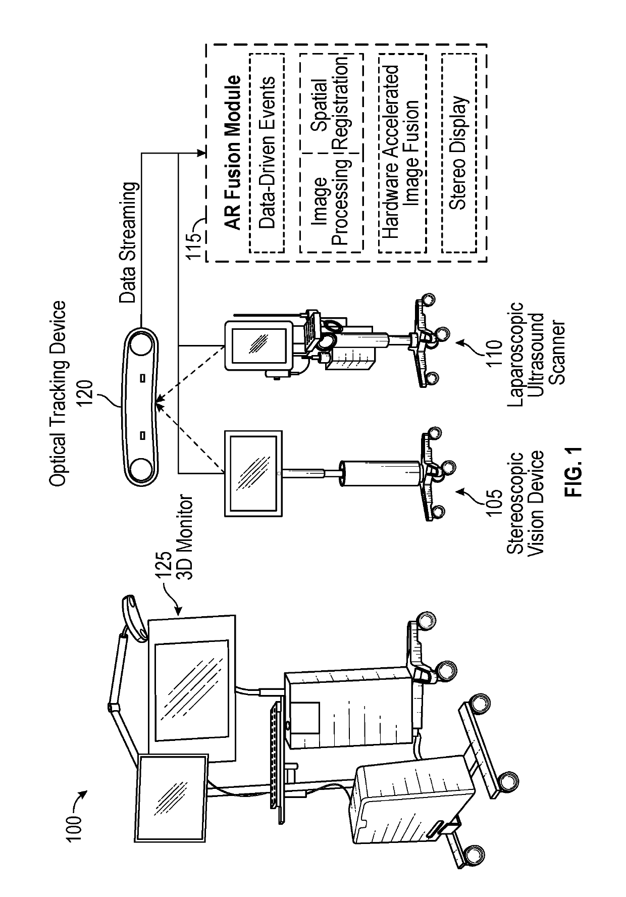
[0060]FIG. 1 illustrates an exemplary stereoscopic AR system and a block diagram AR fusion module with major processing circuitry components for generating composite image data, according to certain embodiments. Stereoscopic AR system 100 includes two imaging devices: a stereoscopic vision device 105 and a laparoscopic ultrasound scanner 110. In certain embodiments, the stereoscopic vision device 105 may be a VSII by Visionsense Corp. with a 5-mm laparoscope (called 3D laparoscope henceforth), and the ultrasound scanner device 110 may be a flex Focus 700 by BK Medical with a 10-mm laparoscopic transducer. In certain embodiments, the 3D laparoscope may be a zero-degree scope with 70-degree field of view. The 3D laparoscope may have a fixed focal length of 2.95 cm. The 3D laparoscope provides an integrated light source and automatic white balance...


PUM


Abstract

A device for generating composite images of dynamically changing surgical anatomy includes circuitry configured to receive, from an endoscopic imaging device, endoscopic image data. The circuitry is configured to receive, from a tomographic imaging device, intra-operative tomographic image data. The circuitry is configured to receive, from a tracking device, spatial tracking data corresponding to the endoscopic imaging device and the tomographic imaging device. The circuitry is configured to generate real-time dynamically changing composite image data by overlaying, based on the spatial tracking data, the intra-operative tomographic image data on the endoscopic image data. The circuitry is configured to output the composite image data to a display.

Description

CROSS REFERENCE TO RELATED APPLICATIONS[0001]This application claims the benefit of priority of U.S. Patent Application No. 61 / 940,915, filed Feb. 18, 2014, and U.S. Patent Application No. 61 / 808,329, filed Apr. 4, 2013 the entire contents of all are incorporated herein by reference.BACKGROUNDTechnical Field[0002]Among other things, the present disclosure is related to minimally invasive surgeries that are carried out under the guidance of real-time video produced by a flexible or rigid endoscope. The present disclosure pertains specifically to combining multiple representations (e.g., medical images or parameters derived from them) of the surgical anatomy to give minimally invasive surgeons a comprehensive view of the surgical field. Whereas one of the representations may be endoscopic video, other representations may be tomographic images acquired both before and / or during surgery. The present disclosure further pertains to a system and methods that permit creating composite image...

Claims


Application Information

Patent Timeline
Patent Type & Authority Patents(United States)
IPC IPC(8): A61B5/00A61B5/06A61B1/00G06T7/38G06T7/33A61B8/08A61B8/00A61B1/313A61B8/12A61B34/20A61B90/00
CPCA61B5/0035A61B8/4416A61B8/483A61B1/00193A61B1/3132A61B5/0073A61B5/7425G06T7/38G06T7/337A61B1/00009A61B5/066A61B8/587A61B8/4209A61B8/5261A61B1/0005A61B8/4245A61B8/12A61B5/062A61B2090/365G06T2207/30056G06T2207/30084A61B2090/378G06T2207/10136G06T2207/10021G06T2207/10081A61B2034/2055G06T2207/10108G06T2207/10104A61B2090/371A61B8/4254A61B1/000094A61B1/00194
Inventor SHEKHAR, RAJKANG, XINAZIZIAN, MAHDIKANE, TIMOTHYPETERS, CRAIG
Owner CHILDRENS NAT MEDICAL CENT
Why Eureka
Try Eureka