
Method and system for transcranial photoacoustic imaging for guiding skull base surgeries

a technology of transcranial photoacoustic imaging and guiding skull, applied in the field of imaging, can solve the problems of high risk of irreversible neurological damage, severe blood loss, stroke or death, and the potential for vision loss

Active Publication Date: 2020-01-14
THE JOHN HOPKINS UNIV SCHOOL OF MEDICINE
View PDF4 Cites 1 Cited by
  • Summary
  • Abstract
  • Description
  • Claims
  • Application Information

AI Technical Summary

Problems solved by technology

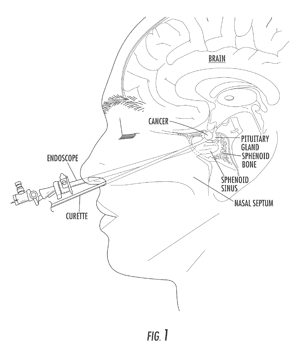
Approximately 8,000 pituitary tumors are diagnosed each year in the United States, causing a variety of hormonal complications, compressing critical nerves and arteries at the base of the brain, and creating a potential for vision loss.
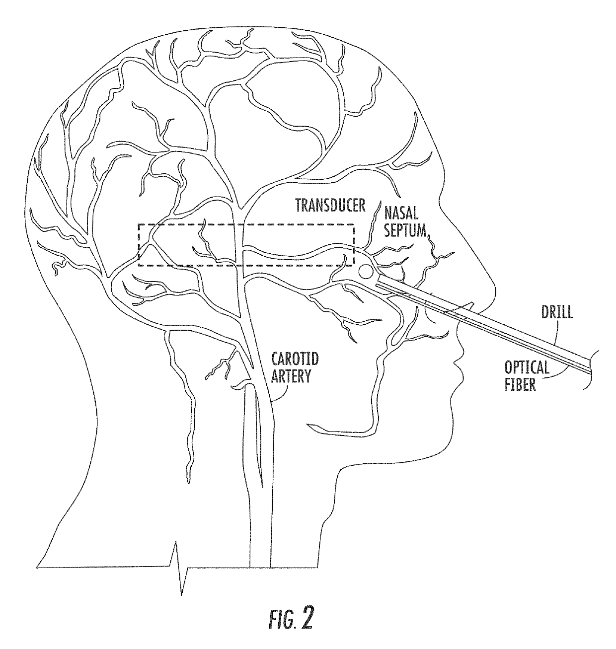
Injury to the carotid arteries, which are located behind the sphenoid on either side of the pea-sized pituitary gland, is a significant complication of transsphenoidal surgery that causes severe blood loss, stroke, or death.
It may be treated with emergency interventions, albeit with a high risk of irreversible neurological damage.
This complication occurs most frequently with novice surgeons who have performed fewer than 200-500 of these surgeries and thus are not sufficiently familiar with potential variations in the anatomy surrounding the pituitary gland.
In addition, this procedure is particularly challenging in pediatric patients who are born with small nasal cavities that mainly develop into their full size after puberty.
Thus, there are generally limited opportunities for novice surgeons to gain necessary caseload experience.
Intraoperative CT may be used for guidance of the bony anatomy surrounding the pituitary tumor, however, it does a poor job of visualizing blood vessels and incurs the risks associated with radiation exposure.
Magnetic resonance angiography is another option, but it is costly and patients with pacemakers or metal implants are not suitable candidates for this approach.
In addition, these imaging modalities are not quite real-time as one volumetric reconstruction could take as long as 20 minutes.
Transcranial ultrasound is a potential option, but it requires low transmit frequencies for skull penetration, which translates to poor spatial resolution and a necessity for expert sonographers to interpret images.
One challenge with conventional photoacoustic imaging methods is the diminishing light penetration, signal-to-noise ratios, and signal contrast as local laser fluence decreases.
This is particularly challenging for transcranial photoacoustic imaging, given the expected poor signal-to-noise ratios due to light obstruction and sound scattering caused by the presence of sphenoidal and temporal bones, respectively.

Method used


Image

  • Method and system for transcranial photoacoustic imaging for guiding skull base surgeries
  • Method and system for transcranial photoacoustic imaging for guiding skull base surgeries
  • Method and system for transcranial photoacoustic imaging for guiding skull base surgeries

Examples


Embodiment Construction

[0062]The presently disclosed subject matter now will be described more fully hereinafter with reference to the accompanying Drawings, in which some, but not all embodiments of the inventions are shown. Like numbers refer to like elements throughout. The presently disclosed subject matter may be embodied in many different forms and should not be construed as limited to the embodiments set forth herein; rather, these embodiments are provided so that this disclosure will satisfy applicable legal requirements. Indeed, many modifications and other embodiments of the presently disclosed subject matter set forth herein will come to mind to one skilled in the art to which the presently disclosed subject matter pertains, having the benefit of the teachings presented in the foregoing descriptions and the associated Drawings. Therefore, it is to be understood that the presently disclosed subject matter is not to be limited to the specific embodiments disclosed and that modifications and other...


PUM


Abstract

The present invention is directed to a method and system for photoacoustic imaging for guiding medical procedures. A transducer is placed near the site of the procedure. The optical fiber, coupled to an electromagnetic source, such as a laser, is attached to a medical device. During the procedure, the device and optical fiber are inserted into the procedure site where the optical fiber illuminates the procedure site, which has a thickness of approximately 2 mm. Photoacoustic images are acquired to visualize the procedure site as the procedure is proceeding in order to provide real-time guidance. This system is applicable to multiple surgical and interventional procedures, such as transsphenoidal surgery.

Description

CROSS REFERENCE TO RELATED APPLICATION[0001]This application claims the benefit of U.S. Provisional Patent Application No. 61 / 934,253 filed on Jan. 31, 2014, which is incorporated by reference, herein, in its entirety.GOVERNMENT SUPPORT[0002]This invention was made with government support under grant number NRI1208540 awarded by the National Science Foundation. The government has certain rights in the invention.FIELD OF THE INVENTION[0003]The present invention relates generally to imaging. More particularly, the present invention relates to a method and system for medical imaging for use in guiding skull base surgeries.BACKGROUND OF THE INVENTION[0004]Approximately 8,000 pituitary tumors are diagnosed each year in the United States, causing a variety of hormonal complications, compressing critical nerves and arteries at the base of the brain, and creating a potential for vision loss. Transsphenoidal surgery is the most common method for removal of pituitary tumors to reverse endocri...

Claims


Application Information

Patent Timeline
Patent Type & Authority Patents(United States)
IPC IPC(8): A61B5/00A61B34/20A61B5/06A61B34/30A61B90/00A61B8/08A61B17/00A61B90/30A61B17/34
CPCA61B8/0841A61B34/30A61B90/37A61B5/0095A61B34/20A61B5/489A61B5/6868A61B5/065A61B2017/345A61B2090/378A61B2090/306A61B2505/05A61B2090/363A61B2090/372A61B2017/00725A61B2034/2055Y10S901/41
Inventor BELL, MUYINATUBOCTOR, EMADKAZANZIDES, PETER
Owner THE JOHN HOPKINS UNIV SCHOOL OF MEDICINE
Learn More