Method of performing electron diffraction pattern analysis upon a sample

Active Publication Date: 2020-05-26
OXFORD INSTR NANOTECH TOOLS
View PDF7 Cites 1 Cited by
  • Summary
  • Abstract
  • Description
  • Claims
  • Application Information

AI Technical Summary

Benefits of technology

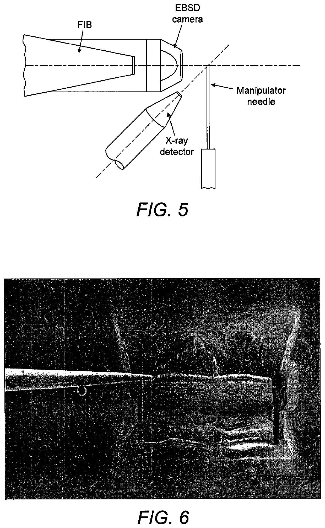
[0015]The invention is performed within the vacuum chamber of analysis apparatus such as a microscope. Thus, the sample is generated from a larger surrounding specimen, is then mounted to the end effector and then oriented into position with respect to the electron beam, all in situ within the apparatus. This removes the need for transferring the sample to a specimen stage by virtue of the “analysis on tip” approach we describe herein.
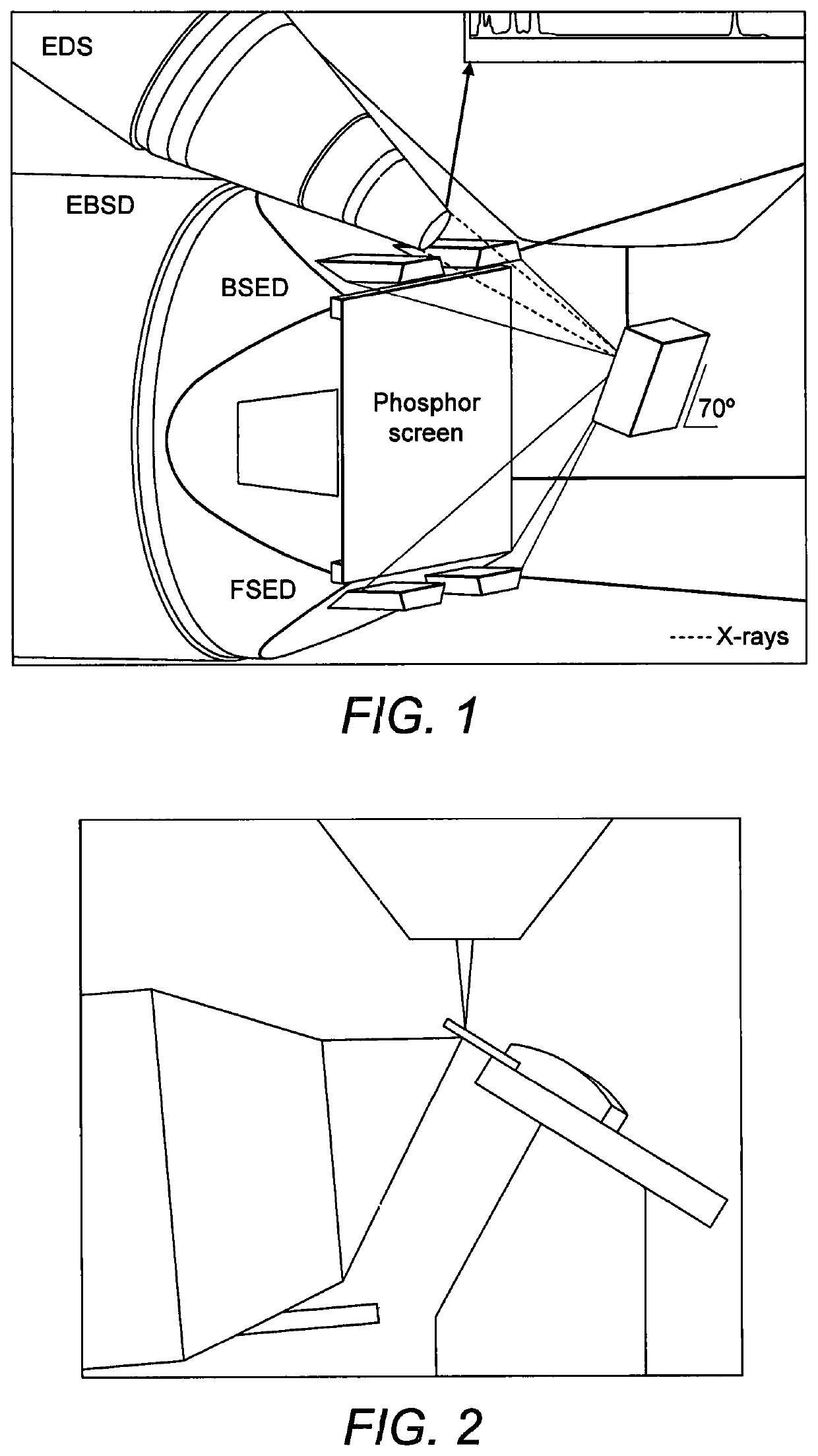
[0026]As will be understood, the few known techniques of performing TKD all rely on elaborate mounting of the sample to the specimen stage of a microscope. These are not needed here. In order to obtain the TKD pattern a camera is positioned so as to view the surface of the sample at a location where the incident electron beam emerges after passing through the sample. This view is therefore of the “underside” of the sample whereas the electron beam impinges upon the “topside” in this case. The present invention is also advantageous in that it allows conventional EBSD to be performed, that is using a camera viewing the surface of the sample at a location where the incident electron beam strikes the sample. In this case the “topside” is viewed by the camera. Where a TKD pattern is to be obtained, the thickness of the sample (distance between the surfaces) is 200 nm or less, preferably 100 nm or less.
[0028]Once the electron diffraction pattern has been obtained (of either type) an analysis of the electron diffraction pattern is used to determine crystalline properties of the material. Typically such analysis is computer-implemented. Such analysis may alone provide the information required by a user. However, a further advantage of the use of the end effector to hold the sample is that an X-ray spectrum may be acquired in the same geometry and from a common overlapping region of the sample as was used to collect the electron diffraction pattern. The X-ray detector only has to have a line of site view of the small volume of material excited by the incident beam and with the sample suspended on the end effector there is less likelihood of occlusion by the stage or bulk specimen from which the sample was extracted. If the sample is in the form or a lamella then there will be a good correlation between the region of material which originates the electron diffraction pattern and that which emits the X-rays. The phase of the material in such as region may then be determined using the analysed crystalline properties of the material (from the electron diffraction analysis) in conjunction with chemical elemental information from analysis of the X-ray spectrum.

Problems solved by technology

Obtaining of a diffraction pattern from a sample whilst mounted to an end effector presents particular challenges regarding the respective orientation needed together with the use of an imaging detector such as a camera.
However the provision of such a connection region is not essential since the sample may be completely isolated prior to attachment of the end effector.

Method used


Image

  • Method of performing electron diffraction pattern analysis upon a sample
  • Method of performing electron diffraction pattern analysis upon a sample
  • Method of performing electron diffraction pattern analysis upon a sample

Examples


Embodiment Construction

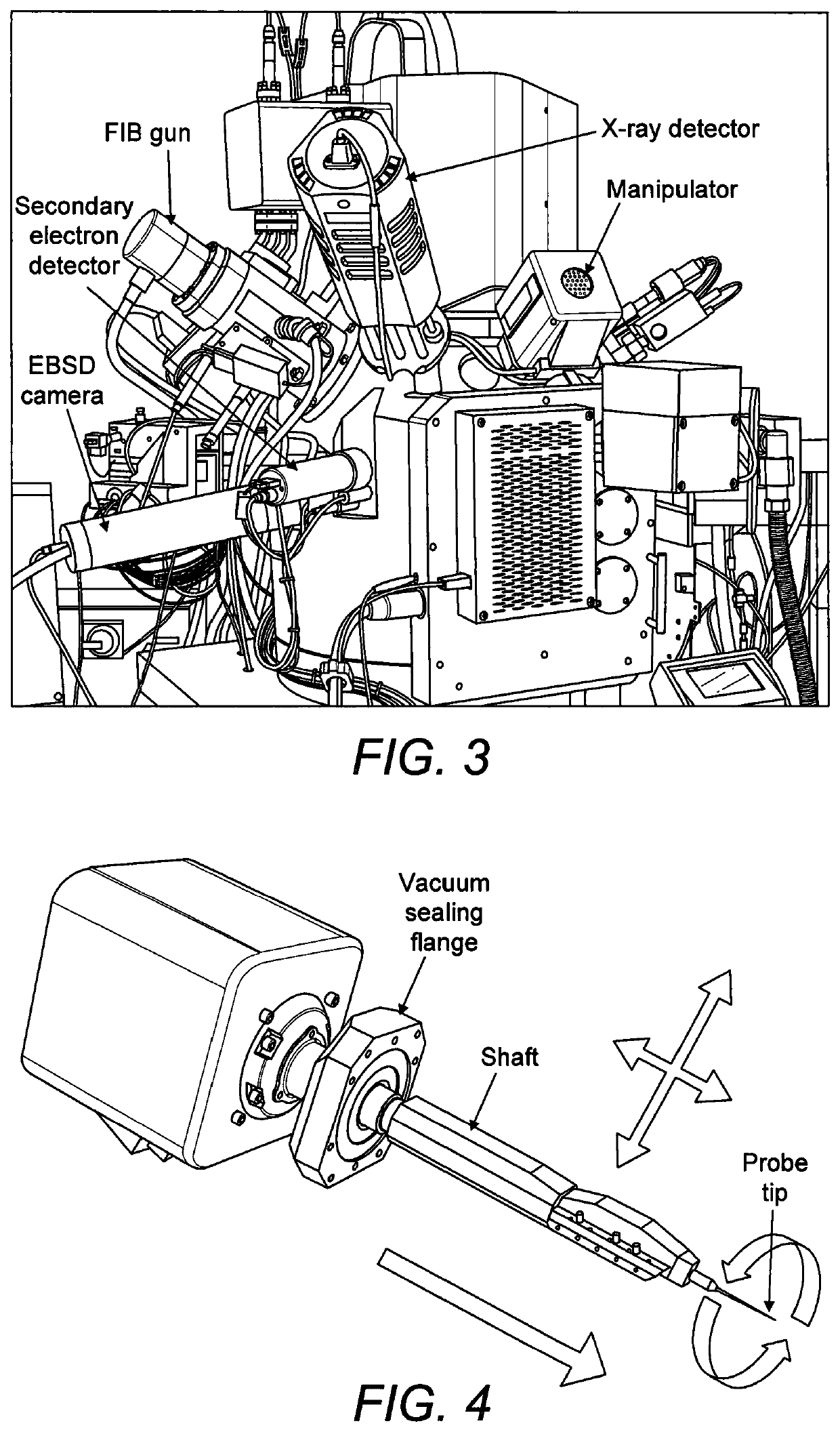
[0048]We now describe example methods effecting a procedure which, while in the vacuum chamber, extracts from the main specimen a small sample that contains the feature of interest and fixes it to the end of a probe that has a controllable rotation about an axis. The probe is arranged to be in a specific orientation with respect to a surface of the sample to be analysed. Thereafter electron diffraction analysis is performed on the sample held by the manipulator. Firstly we describe some practical points associated with the general method.

[0049]The initial procedure of isolation of the sample from the larger specimen is preferably achieved by milling using a focussed particle beam that could be a laser beam or a focussed ion beam (FIB). Using the “FIB lift-out” procedure, FIB milling may be used to isolate a small sample of material from the specimen, attach it to a manipulator end effector (probe) and lift it clear of the specimen. In particular, the FIB may be used to mill a trench...


PUM

PropertyMeasurementUnit
angleaaaaaaaaaa
thicknessaaaaaaaaaa
angleaaaaaaaaaa

Abstract

A method is provided for performing electron diffraction pattern analysis upon a sample in a vacuum chamber of a microscope. Firstly a sample is isolated from part of a specimen using a focused particle beam. A manipulator end effector is then attached to the sample so as to effect a predetermined orientation between the end effector and the sample. With the sample detached, the manipulator end effector is rotated about a rotation axis to bring the sample into a predetermined geometry with respect to an electron beam and diffraction pattern imaging apparatus so as to enable an electron diffraction pattern to be obtained from the sample while the sample is still fixed to the manipulator end effector. An electron beam is caused to impinge upon the sample attached to the manipulator end effector so as to obtain an electron diffraction pattern.

Description

RELATED APPLICATIONS[0001]This application is a national phase of PCT / GB2014 / 050700, filed on Mar. 10, 2014, which claims the benefit of GB Application No. 1402318.8, filed Feb. 11, 2014. The contents of these applications are hereby incorporated by reference.FIELD OF THE INVENTION[0002]The present invention provides a method of performing electron diffraction pattern analysis upon a sample. The method is performed in situ within a vacuum chamber of apparatus such as a focussed ion beam scanning electron microscope.BACKGROUND TO THE INVENTION[0003]In a scanning electron microscope (SEM) a focussed electron beam is rastered over the surface of a specimen. At each point on the raster, an electron detector is used to record the varying signal due to electrons that have been scattered or emitted from the specimen and thus form an image of the rastered field of view. When a small object or feature, such as a particle or an inclusion, is present on the surface of a specimen and this featu...

Claims


Application Information

Patent Timeline
Patent Type & Authority Patents(United States)
IPC IPC(8): G01N23/20058H01J37/295G01N23/2055H01J37/20H01J37/252G01N23/20025G01N23/203H01J37/305
CPCG01N23/2055G01N23/20058G01N23/20025H01J37/20H01J37/252H01J37/3056H01J37/2955G01N23/203H01J2237/31749H01J2237/20221H01J2237/2544H01J2237/20214H01J2237/20207H01J2237/24578
Inventor BAUER, FRANK WILLI
Owner OXFORD INSTR NANOTECH TOOLS
Why Eureka
Try Eureka