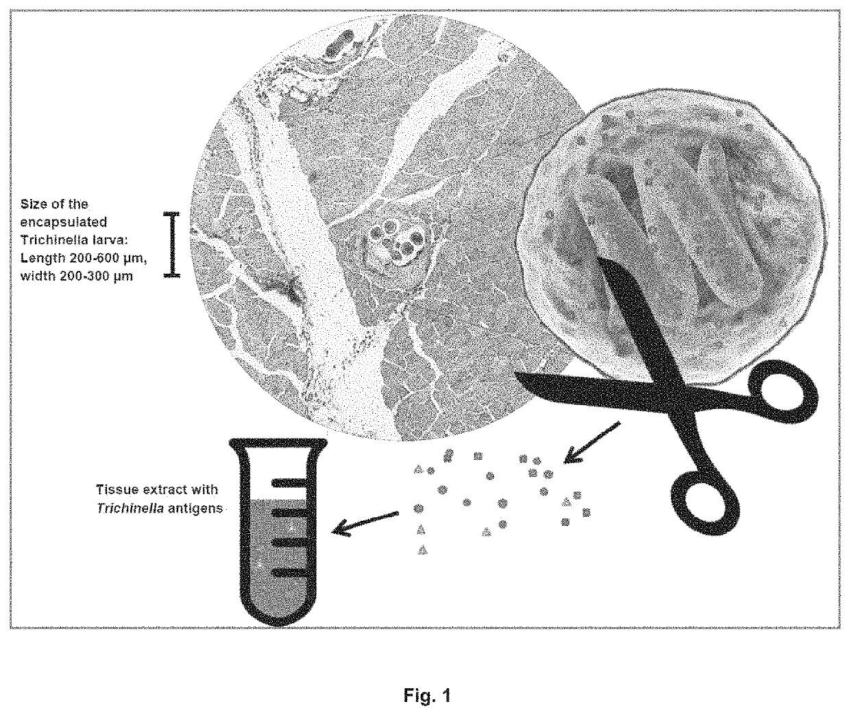
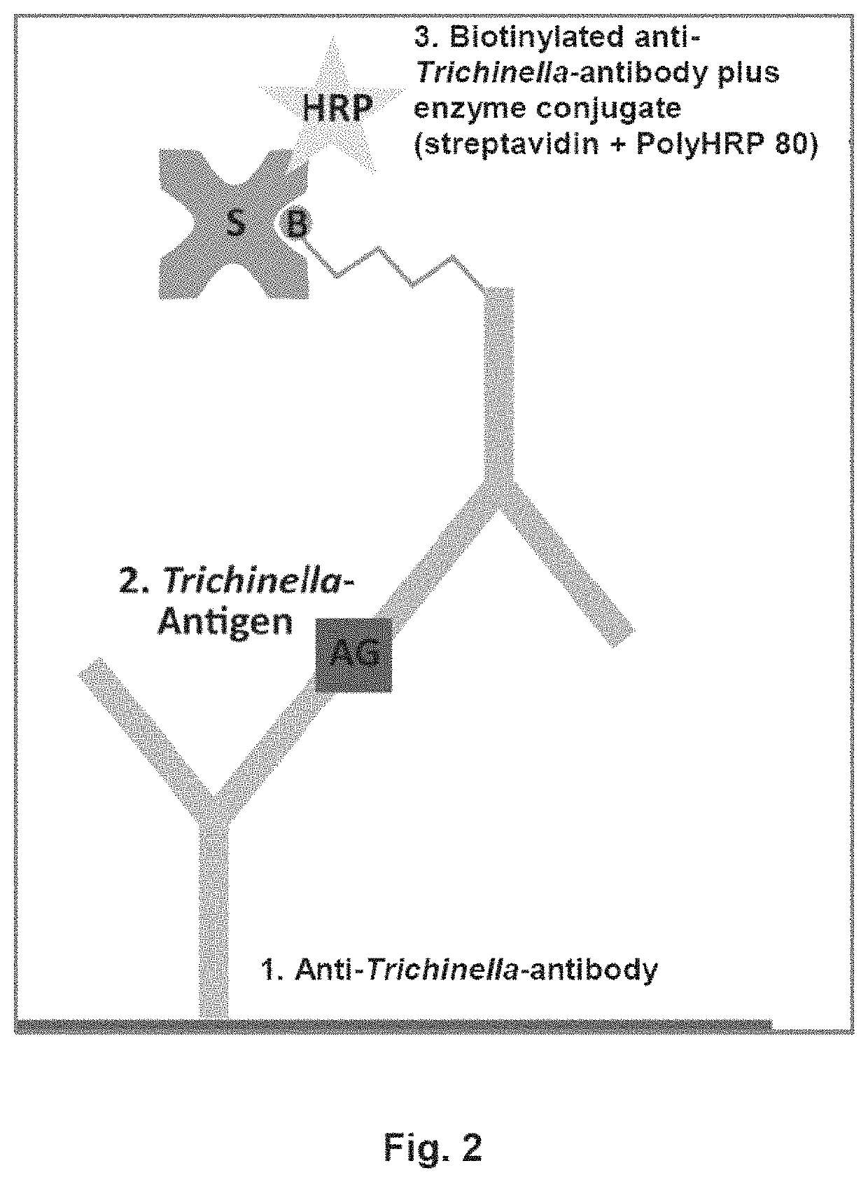
Antigen detection of Trichinella

a trichinella and antigen technology, applied in the field of detecting trichinella, can solve the problems of varying the quality of the pepsin used, temperature and time sensitivity, and serious human diseases, and achieve the effect of reducing the width-to-length ratio

Active Publication Date: 2020-05-26
EUROIMMUN MEDIZINISCHE LABORDIAGNOSTIKA
View PDF5 Cites 0 Cited by
  • Summary
  • Abstract
  • Description
  • Claims
  • Application Information

AI Technical Summary

Problems solved by technology

Trichinella spiralis (T. spiralis) is a nematode of the genus Trichinella and can cause serious human diseases.
Serology is not useful in the Trichina examination, as seroconversion takes place at an infectious dose of 20,000 larvae after 3-4 weeks and of 100 larvae after 5-7 weeks [6].
This method has numerous disadvantages, such as varying quality of the pepsin used, temperature and time sensitivity (digestion should take place for 30 minutes at 46° C. to 48° C.)
, great time intensity (due to the digestion process, sedimentation steps, equipment cleaning and microscopy), manual evaluation by specially trained personnel, difficult and sometimes dangerous handling of individual steps due to working with hydrochloric acid, for example, difficult evaluation (for example, the digestive fluid may have been washed insufficiently and the larvae may thus be overlooked due to the excessive turbidity), and the risk of contamination due to poorly cleaned equipment.
For example, the method mentioned here also has the above-mentioned disadvantages in terms of digestion, since the digestion step is identical.
Furthermore, the sensitivity of the test can be severely impaired due to chemical products such as detergents in cleaning solutions.
In addition, there are many devices that require thorough cleaning, and therefore the risk of contamination is quite high.

Method used


Image

  • Antigen detection of <i>Trichinella</i>
  • Antigen detection of <i>Trichinella</i>
  • Antigen detection of <i>Trichinella</i>

Examples


example 1

and Methods

Material

[0119]

MaterialNameProduct numberManufacturerEquipmentAutomatedPerkinElmerChemiluminescent AnalyzerSuperFlexBiometra WT 15 042-400BiometrashakerCentro XS3 LB 960Berthold TechnologiesMicroplate LuminometerGM 200 Accessories:03 045 0050RetschGrinding container, stainlesssteelGM 200 Accessories: Knife,02 446 0057RetschserratedHydroFlex ™ MicroplateTecanWasher, Magnetic BeadsIncubator WTBBINDERKnife mill GRINDOMIX GM20 253 0001Retsch200MTS 2 / 4 digital microtiter3208001IKAshakerNovex Mini CellInvitrogenPhotometer: Sunrise ™TecanPower Pac HVBio-RadTable centrifuge: BiofugeHeraeusFrescoWasher ColumbusTecanXCell SureLock ™ Mini-CellThermoFisher ScientificElectrophoresis SystemZentrifuge Avanti ® J-EBeckman Coulter(Rotor: JA-10)Tilt / roll mixerIKAConsumablesiD PAGE Gel, 4-20%ID-PA4201-015EurogentecDynabeads ™ M-280 Tosyl-14203ThermoFisher ScientificactivatedMicrotiter plate LockWell446475-EURThermoFisher ScientificMaxisorbNitrocellulose membrane11306SartoriusNunc ™ F96 Micro...

example 2

Comminution of the Meat Samples

[0138]Immediately following comminution of the pork using the GM200 knife mill, particle size determination was carried out using the Camsizer® XT and the HORIBA LA-960. Sampling took place after 6 or 9 min, respectively. The results of the mass distribution can be seen in FIG. 3, where the particle size [μm] is plotted against Q3 [%] in a diagram. Q3 is the percentage of particles that are smaller than x with reference to the total volume. Since the two particle size measuring devices are based on different measuring methods, the results differ.

[0139]The results of the measurement with the HORIBA LA-960 show that 95% of the particles are <26 μm and all particles are <100 μm. With the Camsizer® XT measurement, 95% of the particles are <300 μm. Only 70% of the meat particles are <100 μm. FIG. 4 shows, as an example, what shape the meat particles have. Due to the muscle fibers and myofibrils, the structure is very fibrous, which clearly lowers the width ...

example 3

etection by Means of a Manually Incubated and Automated Chemiluminescence Immunoassay

[0151]For the manually incubated and bead-based chemiluminescence immunoassay (CLIA), the capture antibodies were coupled to magnetic beads rather than to a microtiter plate. Tosyl-activated Dynabeads' were used for this purpose. The hydrophobic polyurethane surface was activated with tosyl groups, which allowed the antibodies to be covalently bound to the beads. 4 mg of the beads were equilibrated with 1 ml of a 1 M Tris buffer. 30 mM Tris, 0.4 M ammonium sulfate, and 28 μg anti-[TRISP][18H1] antibody (abbreviated as Ab 18H1) were added to the equilibrated beads. The incubation was carried out overnight at 37° C., on a roller mixer. The next day, the beads were washed three times with 1 ml each of StabilCoat® Plus, and then incubated overnight at 37° C., on the roller mixer, to block any remaining reactive functional groups. After blocking, the beads were taken up in fresh StabilCoat® Plus and stor...


PUM

PropertyMeasurementUnit
diameteraaaaaaaaaa
diameteraaaaaaaaaa
diameteraaaaaaaaaa

Abstract

Trichinella can be detected in a tissue extract sample. A suitable kit includes (a) a detection carrier containing a first antibody against one or more antigens of Trichinella, and (b) (i) a second antibody against one or more antigens of Trichinella, wherein the second antibody is bound to a signal molecule, or (b) (ii) contains one or more antigens of Trichinella bound to a signal molecule, wherein the antigens of (b) (ii) are configured so as to dissolve binding of the antigens of (a) to the first antibody by competitive displacement.

Description

CROSS-REFERENCE TO RELATED APPLICATIONS[0001]The present application claims priority to European patent applications EP 17 208 994.8 filed Dec. 20, 2017 and EP 18 199 404.7 filed Oct. 9, 2018, the content of each of which is incorporated by reference in its entirety.REFERENCE TO A SEQUENCE LISTING[0002]The instant application contains a Sequence Listing which has been filed electronically in ASCII format and is hereby incorporated by reference in its entirety. Said ASCII copy, created on Dec. 18, 2018, is named 181203_Sequence_Listing-as-filed.txt and is 6,692 bytes in size.BACKGROUND OF THE INVENTIONField of the Invention[0003]The present invention is directed at a method of detecting Trichinella in a tissue extract sample, the use of a detection system according to the invention for detection of Trichinella, and a kit comprising (a) a detection carrier comprising a first antibody against one or more antigens of Trichinella, and (b) (i) a second antibody against one or more antigen...

Claims


Application Information

Patent Timeline
Patent Type & Authority Patents(United States)
IPC IPC(8): G01N33/53G01N33/487G01N33/569G01N33/12G01N33/68C07K16/18G01N33/58C07K14/435
CPCG01N33/48735G01N33/12G01N33/569C07K14/4354C07K16/18G01N33/5308G01N33/6854G01N33/58G01N2333/4353G01N33/53A61K39/00C12N1/20
Inventor BRAASCH, JANAOSTERMANN, STEFANIEMACKIEWICZ, MONIKA
Owner EUROIMMUN MEDIZINISCHE LABORDIAGNOSTIKA
Why Eureka
Try Eureka