Spatial molecular analysis of tissue

a tissue and molecular analysis technology, applied in the field of spatial molecular analysis of tissue, can solve the problems of inability to maintain spatial information, low throughput and laborious constraints, and achieve the effect of avoiding cross-talk, significant functional benefits, and reliable and efficient tissue molecular analysis

Active Publication Date: 2020-07-28
MAYO FOUND FOR MEDICAL EDUCATION & RES +1
View PDF12 Cites 2 Cited by
  • Summary
  • Abstract
  • Description
  • Claims
  • Application Information

AI Technical Summary

Benefits of technology

[0006]The methods and systems provided herein overcome conventional limitations and problems associated with spatial imaging of tissue samples, including spatial gene expression useful for tissue characterization. The ability to reliably and efficiently achieve spatial molecular analysis of tissue relies on pixelating tissue samples into individual wells. Corresponding efficient processing of the pixelated tissue, including by reliable interaction of reagent materials and tissue, bulk fluid application and removal, and avoiding cross-talk between different wells containing different pixelated tissue, together ensure the methods and systems provide significant functional benefits that ensure reliable spatial mapping of a tissue sample.
[0014]The deformable layer may comprise a polymer or an elastomer, or any material that exhibits a deformation property such that after the applied force is removed, the deformable layer exits the wells and relaxes back to a rest state. In contrast, the plurality of tissue sample islands remains within the wells. The wells may be coated with an adhesion-promoting layer that ensures a bonding force between the tissue and the well that is greater than the adhesion force between the tissue and the deformable layer. This ensures that tissue islands remain in the wells even when the deformable layer exits the wells. Accordingly, the deformable layer may be coated with an anti-adhesion-promoting layer to minimize the adhesive force between the tissue and the deformable substrate.
[0018]The method may further comprise the step of processing the tissue sample islands by: removing the deformable substrate; applying reagents used to image and / or quantify the tissue sample islands to each of the wells, wherein the applying step comprises: covering the wells with liquid reagent, wherein the liquid reagent enters the wells by capillary action; immersing the wells with liquid reagent in an inert covering fluid having a density that is less than the liquid reagent density, thereby enveloping each well containing a tissue sample island and liquid reagent without entering the wells; and removing excess reagent by forcing a gas over the microarray, thereby avoiding cross-talk between different wells. In this manner, the liquid filling is rapid, reliable, and avoids unwanted material communication between adjacent wells. The covering fluid may comprise mineral oil.
[0032]Also provided herein is a method for generating a pixelated, spatially-preserved tissue sample. Numerous functional advantages are achieved with such methods, including the ability to rapidly, reliably and at a high sensitivity and resolution, characterize molecular variation of a tissue sample, The method may comprise the steps of: providing a microarray having a plurality of wells, wherein at least a portion of each edge of the well is a shearing surface; providing a tissue sample in contact with each of the wells; overlaying a deformable layer on said tissue sample; applying a force upon the deformable layer, thereby forcing the deformable layer and the tissue sample into the plurality of wells and shearing the tissue sample into a plurality of tissue sample islands positioned in the plurality of wells; and relaxing the force, thereby removing the deformable layer from the plurality of wells, while maintaining the plurality of tissue sample islands positioned in the plurality of wells, thereby generating a pixelated, spatially-preserved tissue sample.
[0038]Any of the methods described herein may have a plurality of PCR products that are fluorescent, thereby facilitating imaging and, if desired, quantifying.

Problems solved by technology

The spatial localization of gene expression can unravel important insights into tissue heterogeneity, functionality and pathological transformations, but the ability to maintain this spatial information remains an enduring challenge in tissue sections routinely used for pathology.
Amplification-based spatial gene expression analysis methods provide good sensitivity and specificity but decouple the analyte isolation and biochemical detection steps, making them low throughput and laborious constraints, limiting the translation of the above methods into routine research and clinical practice.
In addition, off-target binding of FISH probes and cellular auto-fluorescence can also become a limiting factor in imaging tissue samples.

Method used


Image

  • Spatial molecular analysis of tissue
  • Spatial molecular analysis of tissue
  • Spatial molecular analysis of tissue

Examples


example 1

nalysis of Prostate Cancer

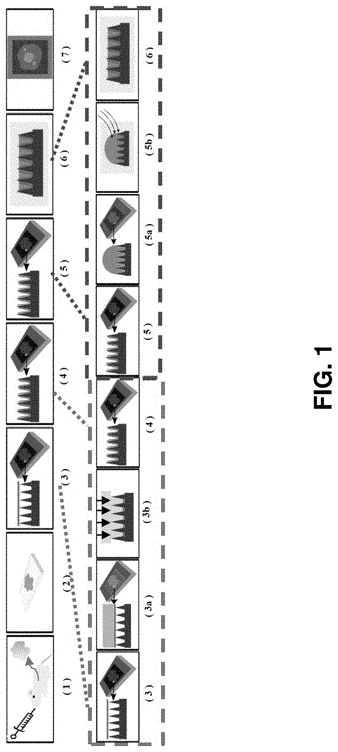
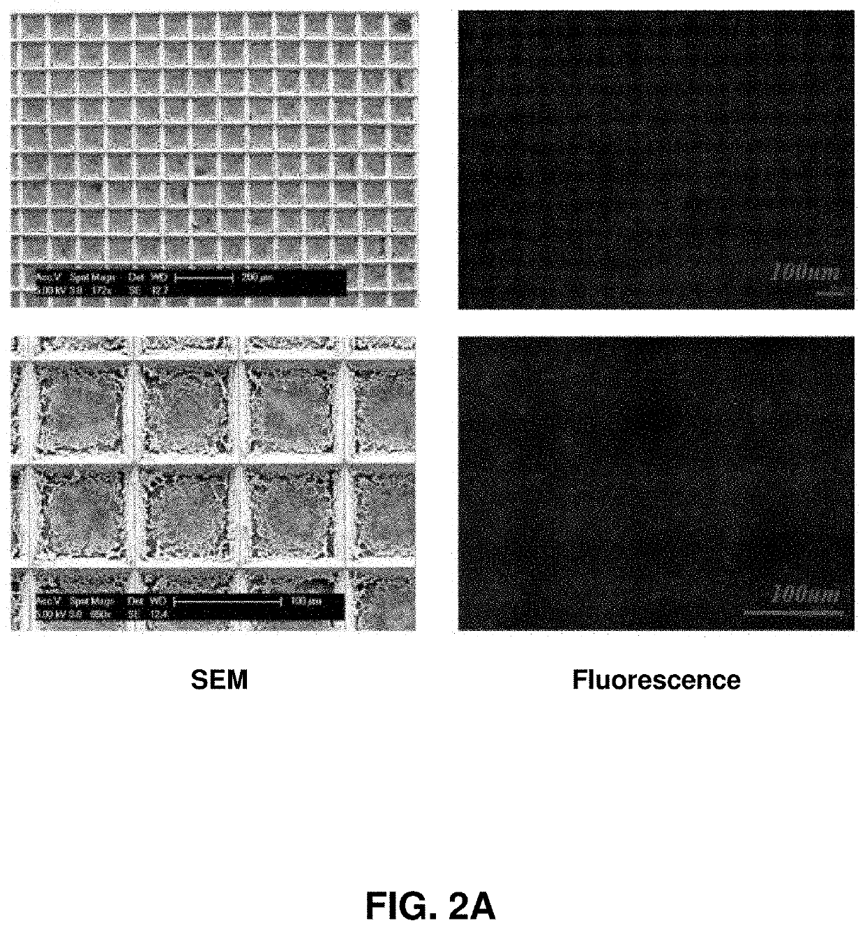
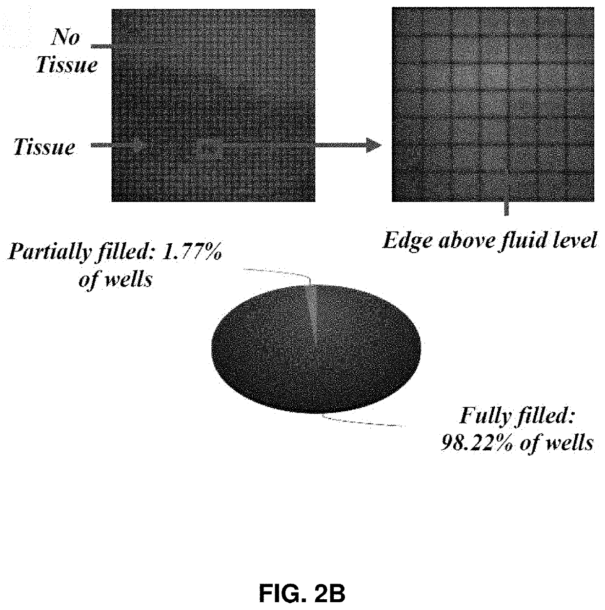
[0090]Described herein is an on-chip spatial gene expression analysis technique that can perform real-time nucleic acid amplification, including reverse transcriptase loop mediated isothermal amplification (RT-LAMP) starting from tissue samples, while keeping the native spatial location of the nucleic acid preserved. We engineered a silicon oxide chip with an array of microwells that serve as independent picoliter volume RT-LAMP reaction vessels. The wells were designed to have knife-like sharp edges (referred herein as a “shearing surface”) that help in tissue partitioning-and-transfer into wells starting from a tissue cryosection. A capillary action based reagent loading technique was developed to fill all the wells on the chip simultaneously while preventing reagent overflow between wells in the final loaded chip. Using this platform we amplified the TOP2A mRNA starting from a 7 micron tissue section of a prostate cancer xenograft and visualized the vari...

example 2

Spatial Gene Expression Analysis from Tissue

[0110]Described herein is a rapid technique that performs on-chip picoliter real-time reverse transcriptase loop mediated isothermal amplification (RT-LAMP) reactions on a histological tissue section without any analyte purification while maintaining the native spatial location of the nucleic acid molecules. This example includes a method of amplifying TOP2A messenger RNA (mRNA) in a prostate cancer xenograft with 100 μm spatial resolution and by visualizing the variation in threshold times across the tissue. The on-chip reaction was validated through fluorescence mRNA in situ hybridization (ISH). The entire process, from tissue loading on microchip to results from RT-LAMP can be carried out in less than two hours. This technique with its ease of use, fast turnaround, and quantitative outputs is an invaluable tissue analysis tool for researchers and clinicians in the biomedical arena.

[0111]The spatial localization of gene expression can un...

example 3

r Generating a Pixelized Tissue Sample

[0189]In an embodiment, described herein is a device for pixelating a tissue sample. An exemplary device is provided in FIGS. 22A-K and 31A-B. The device includes a plurality of wells 100 supported by or embedded in a substrate 110 for receiving a tissue sample, including a cryogenic histogram. A shearing surface 120 positioned between the wells has a sharp edge to sever a tissue sample 120 when force is applied to it. A deformable substrate 140 configured to fit over the plurality of wells 100 and placed on top of the tissue sample may be used to force tissue sample into the wells and ensure the tissue sample 130 is severed such that a portion (e.g., “island”) enters each of the plurality of wells 100. When force, such as that provided by a centrifuge, is applied, as indicated by the arrow in FIG. 31B, the tissue sample severs such that a portion is forced into each of the wells generating a tissue sample island 150 in each well and thereby cre...


PUM


Abstract

Various methods and devices for spatial molecular analysis from tissue is provided. For example, a method of spatially mapping a tissue sample is provided with a microarray having a plurality of wells, wherein adjacent wells are separated by a shearing surface; overlaying said microarray with a tissue sample; applying a deformable substrate to an upper surface of said tissue sample; applying a force to the deformable substrate, thereby forcing underlying tissue sample into the plurality of wells; shearing the tissue sample along the shearing surface into a plurality of tissue sample islands, with each unique tissue sample island positioned in a unique well; and imaging or quantifying said plurality of tissue sample islands, thereby generating a spatial map of said tissue sample. The imaging and / or quantifying may use a nucleic acid amplification technique.

Description

CROSS-REFERENCE TO RELATED APPLICATIONS[0001]This application claims the benefit of and priority to U.S. Provisional Application No. 62 / 404,825, filed Oct. 6, 2016, which is hereby incorporated in its entirety to the extent not inconsistent herewith.STATEMENT REGARDING FEDERALLY SPONSORED RESEARCH OR DEVELOPMENT[0002]This invention was made with government support under 1534126 awarded by The National Science Foundation, 59-8072-6-001 awarded by The United States Department of Agriculture, and 087126 awarded by The National Institutes of Health. The government has certain rights in the invention.REFERENCE TO A SEQUENCE LISTING[0003]A sequence listing containing SEQ. ID. NOs. 1-44 is submitted herewith and is specifically incorporated by reference.BACKGROUND OF INVENTION[0004]The spatial localization of gene expression can unravel important insights into tissue heterogeneity, functionality and pathological transformations, but the ability to maintain this spatial information remains ...

Claims


Application Information

Patent Timeline
Patent Type & Authority Patents(United States)
IPC IPC(8): C12P19/34B01L3/00C12N15/10C12Q1/6874C12Q1/686G16B25/10G16B25/00G01N1/28G16B30/00C12Q1/6827
CPCC12Q1/6874C12N15/1003G16B25/00B01L3/5085C12Q1/686B01L3/5027C12N15/1096G01N1/286G16B25/10C12Q2521/107B01L2300/0893G16B30/00B01L2300/0858G01N2001/2873B01L2200/0642B01L3/50851B01L2300/0819C12Q1/6827G01N2001/282
Inventor BASHIR, RASHIDGANGULI, ANURUPKOSARI, FARHAD
Owner MAYO FOUND FOR MEDICAL EDUCATION & RES
Why Eureka
Try Eureka