

Methods for lymphatic delivery of active agents
a technology of active agents and lymph nodes, applied in the direction of infusion needles, drug compositions, immunological disorders, etc., can solve the problems of ineffective targeting and direct delivery of drugs to the lymphatic system, lymph node swelling or lymphadenitis, and orally delivered drugs often suffer from first-pass metabolism effects, etc., to reduce the symptoms of disease, delay the progression of the disease, and treat the effect of diseas
- Summary
- Abstract
- Description
- Claims
- Application Information
AI Technical Summary
Benefits of technology
Problems solved by technology
Method used
Image

Examples
example 1
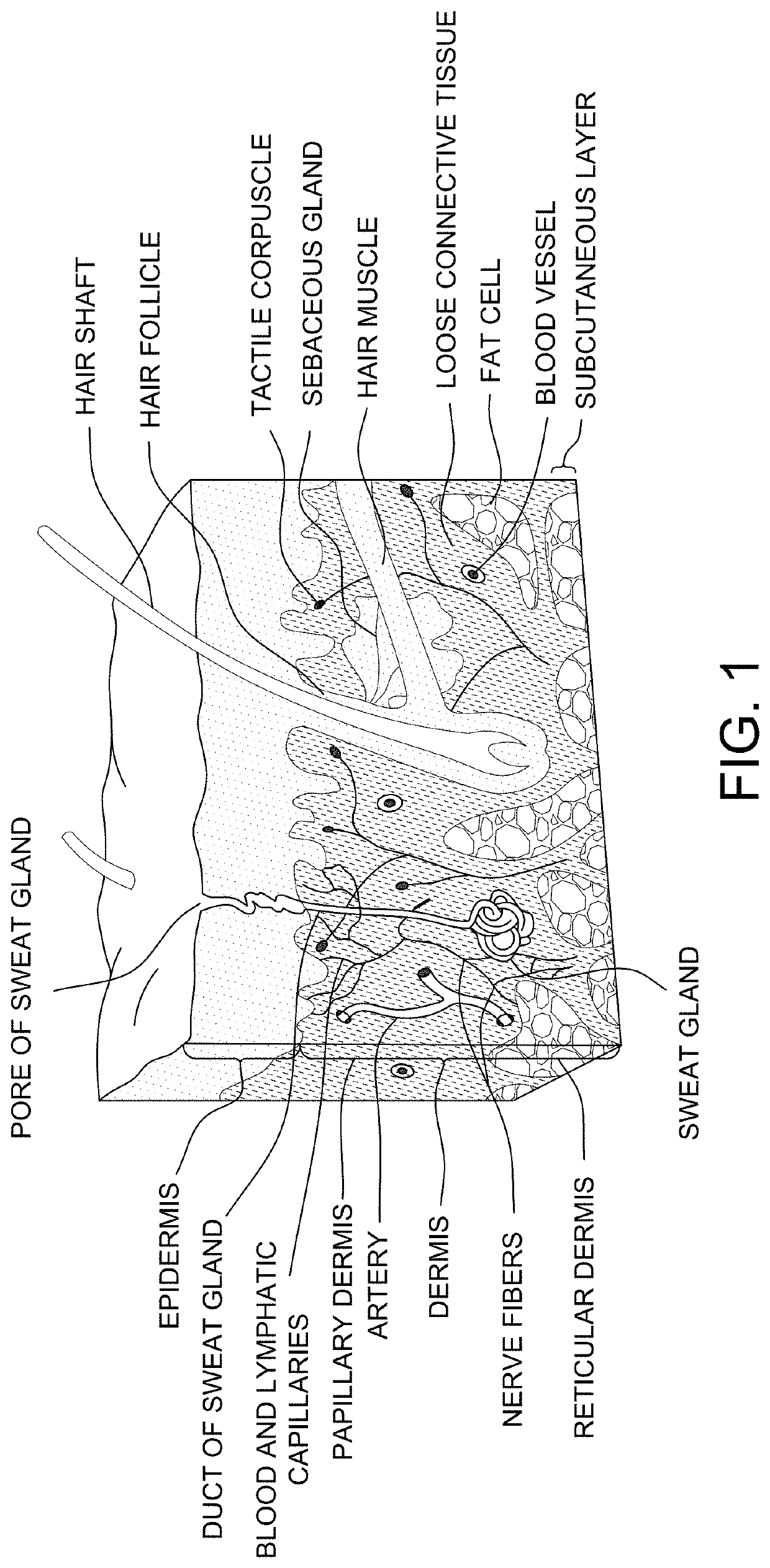
Diagram of the Skin
[0176]The overall structure of the skin including the dermis and epidermis is illustrated in FIG. 1. The dermis is composed of a myriad of tissue types, and in general, exhibits an overall thickness ranging from about 500 μm to about 4,000 μm. The lymphatic and blood capillaries are found often found together as illustrated or they can be present as separate entities. As illustrated, the blood and lymphatic capillaries are often located within the upper portions of the dermis (e.g., near the epidermal dermal or epidermal basement membrane) within a portion of the papillary dermis. Larger vessels are generally found within the lower reticular dermis (e.g., a blood vessel as shown). Other tissue types important to dermal function include the larger arteries, arterioles, sweat gland ducts, sebaceous glands, nerve corpuscles, connective tissues and extra cellular matricis, smooth muscle, and hair follicles. Below the reticular dermis lies the subcutaneous tissue layer...
example 2
Depth of Dye Penetration into the Skin
[0178]An array of needles was fabricated on a patch and was used to estimate the average range of depth of delivering of an agent within the skin of adult guinea pigs. As shown in FIG. 5, methylene blue dye was administered to an average depth of about 92 μm demonstrating a range of depth distributions of about 5 μm to about 200 μm. As shown in FIG. 6, the structure and depth in the skin may be estimated by using optical coherence tomography techniques. The structure of the skin after applying an array of needles can be visualized by looking at individual horizontal slices of the skin.
example 3
Modulation of Epidermal Tight Junction Proteins
[0179]An array of needles having a nanotopography surface was fabricated on a patch and tested on an in vitro mono layer of Caco-2 epithelial cells. As shown in FIG. 7A, the ZO-1 tight junction protein shows a normal staining pattern. However, when an array of needles having a nanotopography surface is placed within proximity of Caco-2 cells, a disrupted staining pattern can be visualized. This ruffled pattern indicates junction remodeling in the areas of where the nanotopography was located (FIG. 7B). When the array of needles having a nanotopography is removed, the staining pattern returns to normal, indicating a spatial and temporal effect on tight junction proteins, such as ZO-1 (FIG. 7C).
PUM
 Abstract
Description
Claims
Application Information