
Anti-myostatin antibodies, polypeptides containing variant Fc regions, and methods of use

a technology of myostatin and polypeptides, which is applied in the field of anti-myostatin antibodies, can solve the problems of limited treatment options for these disorders, increased risk of thromboembolism, so as to reduce body fat accumulation, increase muscle mass, and increase the effect of muscle mass

Active Publication Date: 2020-08-11
CHUGAI PHARMA CO LTD
View PDF476 Cites 10 Cited by
  • Summary
  • Abstract
  • Description
  • Claims
  • Application Information

AI Technical Summary

Benefits of technology

The present invention is about anti-myostatin antibodies that can be used as a medicament to treat muscle wasting diseases, increase muscle tissue mass and strength, or reduce body fat accumulation. The technical effect of this patent is the discovery of these antibodies and their potential use in treating muscle wasting diseases and promoting muscle tissue growth and strength.

Problems solved by technology

Patients will benefit from an increase in muscle mass and / or muscle strength; however, there are presently limited treatments available for these disorders.
However, these prior art antibodies are all specific for mature myostatin but not for latent myostatin, and the strategies described for inhibiting myostatin activity have utilized antibodies that can bind to and neutralize mature myostatin.
For example, the group of patients who were administered bevacizumab, an antibody against VEGF, is known to have an increased risk for thromboembolism (see, e.g., Scappaticci et al., J. Natl. Cancer. Inst.
Administering an antibody with enhanced FcγRIIa binding to such patients who already have a high risk for developing thromboembolism will increase the risk for developing thromboembolism, thus is extremely dangerous.
When antibodies are administered as pharmaceuticals, it is supposed that peptide fragments derived from the administered antibodies are likely to be also presented as an antigen, thereby increasing the risk of production of antibodies against therapeutic antibodies (anti-therapeutic antibodies).
More specifically, enhancing FcγRIIa binding will increase the risk of production of antibodies against the therapeutic antibodies, and this will remarkably decrease their value as pharmaceuticals.
More specifically, the value as pharmaceuticals will be considerably reduced when FcγRIIa binding is enhanced, which leads to increased risk of thrombus formation via platelet aggregation and increased risk of anti-therapeutic antibody production due to an increased immunogenicity.
Therefore, when this Fc variant was used for treatment of autoimmune diseases, the number of patients who can safely use it while enjoying its effects as a pharmaceutical will be limited.
Therefore, it is considered to be difficult for these variants to actually induce an FcγRIIb-mediated immunosuppressive reaction more strongly than IgG1.
However, when FcγRIIa binding is similarly enhanced, unintended activities such as ADCC activity and ADCP activity will be exhibited, and this may cause side effects.
Therefore, the difficult problem may be producing an Fc region variant with enhanced selective FcγRIIb binding as compared to each allotype of FcγRIIa, which involves distinguishing highly homologous sequences between FcγRIIa and FcγRIIb.
Owing to the absence of such an Fc variant, the effects of the Fc variant selectively binding to FcγRIIb have not been thoroughly tested yet in monkey.

Method used


Image

  • Anti-myostatin antibodies, polypeptides containing variant Fc regions, and methods of use
  • Anti-myostatin antibodies, polypeptides containing variant Fc regions, and methods of use
  • Anti-myostatin antibodies, polypeptides containing variant Fc regions, and methods of use

Examples


example 1

Expression and Purification of Human, Cynomolgus Monkey, and Mouse Myostatin Latent and Mature Form

[0587]Human latent myostatin (also described herein as human myostatin latent form) (SEQ ID NO: 1) was expressed transiently using FREESTYLE®293 cells (FS293-F cells) (Thermo Fisher, Carlsbad, Calif., USA). Conditioned media containing expressed human myostatin latent form was acidified to pH6.8 and diluted with ½ vol of milliQ water, followed by application to a Q-sepharose FF anion exchange column (GE healthcare, Uppsala, Sweden). The flow-through fraction was adjusted to pH5.0 and applied to a SP-sepharose HP cation exchange column (GE healthcare, Uppsala, Sweden), and then eluted with a NaCl gradient. Fractions containing the human myostatin latent form were collected and subsequently subjected to a SUPERDEX® 200 gel filtration column (GE healthcare, Uppsala, Sweden) equilibrated with 1×PBS. Fractions containing the human myostatin latent form were then pooled and stored at −80° C....

example 2

Identification of Anti-Latent Myostatin Antibody

[0590]Anti-latent myostatin antibodies were prepared, selected, and assayed as follows.

[0591]Twelve to sixteen week old NZW rabbits were immunized intradermally with mouse latent myostatin and / or human latent myostatin (50-100 μg / dose / rabbit). This dose was repeated 3-4 times over a one month period. One week after the final immunization, the spleen and blood from the immunized rabbit were collected. Antigen-specific B-cells were stained with labelled antigen, sorted with FCM cell sorter (FACS aria III, BD), and plated in 96-well plates at a one cell / well density together with 25,000 cells / well of EL4 cells (European Collection of Cell Cultures) and with rabbit T-cell conditioned medium diluted 20 times, and were cultured for 7-12 days. EL4 cells were treated with mitomycin C (Sigma) for 2 hours and washed 3 times in advance. The rabbit T-cell conditioned medium was prepared by culturing rabbit thymocytes in RPMI-1640 containing Phytoh...

example 3

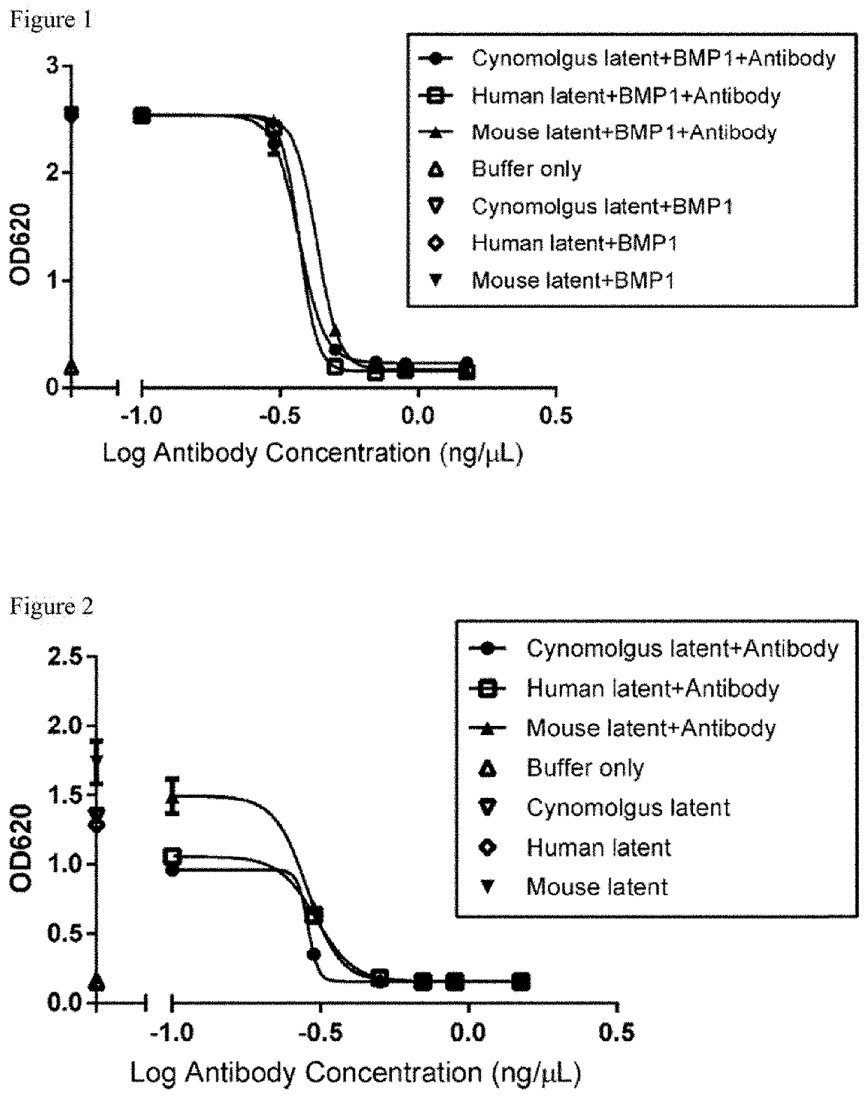
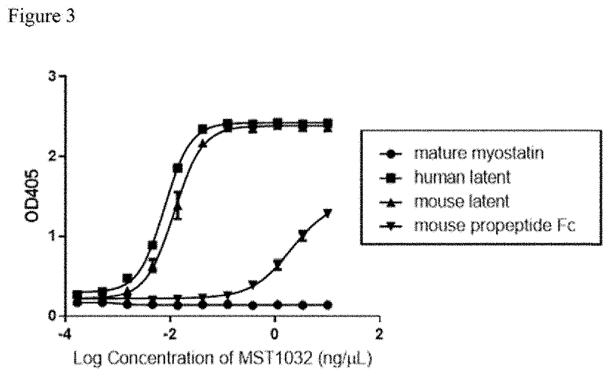
Characterization of Anti-Latent Myostatin Antibody (HEK Blue Assay (BMP1 Activation))

[0595]A reporter gene assay was used to assess the biological activity of active myostatin in vitro. HEK-Blue™ TGF-β cells (Invivogen) which express a Smad3 / 4-binding elements (SBE)-inducible SEAP reporter genes, allow the detection of bioactive myostatin by monitoring the activation of the activin type 1 and type 2 receptors. Active myostatin stimulates the production of SEAP which is secreted into the cell supernatant. The quantity of SEAP secreted is then assessed using QUANTIBlue™ (Invivogen).

[0596]HEK-Blue™ TGF-β cells were maintained in DMEM medium (Gibco) supplemented with 10% fetal bovine serum, 50 μg / mL streptomycin, 50 U / mL penicillin, 100 μg / mL Normocin™, 30 μg / mL of Blasticidin, 200 μg / mL of HygroGold™ and 100 μg / mL of Zeocin™. During the functional assay, cells were changed to assay medium (DMEM with 0.1% bovine serum albumin, streptomycin, penicillin and Normocin™) and seeded to a 96-w...


PUM

PropertyMeasurementUnit
strengthaaaaaaaaaa
massaaaaaaaaaa
lengthaaaaaaaaaa

Abstract

The disclosure provides anti-myostatin antibodies and methods of making and using the same. Nucleic acids encoding the anti-myostatin antibodies and host cells comprising the nucleic acids are also provided. The anti-myostatin antibodies have uses that include treating a muscle wasting disease, reducing body fat accumulation, and increasing mass and strength of muscle tissue. The disclosure also provides polypeptides containing a variant Fc region and methods of making and using the same. Nucleic acids encoding the polypeptides and host cells comprising the nucleic acids are also provided. The polypeptides have uses that include suppressing the activation of immune cells; treating an immunological inflammatory disease, autoimmune disease, or viral infection; and increasing muscle mass and strength or reducing body fat accumulation.

Description

CROSS REFERENCE TO RELATED APPLICATIONS[0001]This application is a divisional of U.S. patent application Ser. No. 14 / 974,488, filed Dec. 18, 2015, now U.S. Pat. No. 10,000,560 B2, issued Jun. 19, 2018, which claims priority to Japanese Patent Application No. 2014-257636, filed Dec. 19, 2014, each of which is incorporated herein by reference.REFERENCE TO SEQUENCE LISTING SUBMITTED ELECTRONICALLY[0002]The content of the electronically submitted sequence listing in ASCII text file (Name: 6663.0064_Sequence_Listing.txt; Size: 660,306 bytes; and Date of Creation: Apr. 16, 2018) filed with the application is herein incorporated by reference in its entirety.BACKGROUND[0003]The present invention relates to anti-myostatin antibodies and methods of using the same. The present invention also relates to polypeptides containing variant Fc regions and methods of using the same. Myostatin, also referred to as growth differentiation factor-8 (GDF8), is a secreted protein and is a member of the tran...

Claims


Application Information

Patent Timeline
Patent Type & Authority Patents(United States)
IPC IPC(8): C07K16/22A61K39/00G01N33/68
CPCG01N33/6872C07K16/22C07K2317/33C07K2317/34C07K2317/92C12N2510/02A61K2039/505G01N2333/495C07K2317/24C07K2317/94C07K2317/52C07K2317/565C07K2317/76A61P1/16A61P11/00A61P13/12A61P21/00A61P21/02A61P21/04A61P35/00A61P43/00A61P7/00A61P9/00A61P9/04C07K2317/14G01N33/6854
Inventor RUIKE, YOSHINAOKURAMOCHI, TAICHIMURAMATSU, HIROYASUUEYAMA, ATSUNORIIGAWA, TOMOYUKIKATADA, HITOSHIHORI, YUJI
Owner CHUGAI PHARMA CO LTD
Learn More