In vitro gastrointestinal model comprising lamina propria-derived cells

a technology of lamina propria and gastrointestinal tract, which is applied in the field of in vitro microfluidic gutonchip, to achieve the effect of reducing inflammation

Active Publication Date: 2021-05-11
EMULATE INC
View PDF18 Cites 1 Cited by
  • Summary
  • Abstract
  • Description
  • Claims
  • Application Information

AI Technical Summary

Benefits of technology

The patent describes a microfluidic gut-on-chip that can mimic the structure and functions of the gastrointestinal system in humans. The chip includes layers of intestinal epithelial cells, primary immune cells, and vascular endothelial cells that work together to recapitulate key aspects of intestinal physiology and pathophysiology. The chip can be used to model inflammatory gastrointestinal tissue, compare different types of gastrointestinal tissue, and identify cells and factors driving disease states. The chip can also be used for drug testing and to identify new treatments for inflammatory gastrointestinal disorders. The use of microfluidic technology allows for the creation of multiple duplicate chips and the co-culturing of primary cells for up to 9 days without significant cell death. The chip has been shown to mimic an ulcerative colitis-like disease phenotype and can be used to identify new treatments for inflammatory gastrointestinal disorders.

Problems solved by technology

However, it is not meant to limit gastrointestinal tissue, gastrointestinal cells, stromal cells, LPDCs, immune cells, or other cell types or tissues from humans.

Method used


Image

  • In vitro gastrointestinal model comprising lamina propria-derived cells
  • In vitro gastrointestinal model comprising lamina propria-derived cells
  • In vitro gastrointestinal model comprising lamina propria-derived cells

Examples


example 1

a Gut-On-Chip

[0543]The following exemplary protocol outlines the sterilization / functionalization, coating, media wash, and cell seeding steps for preparing the Intestine-on-Chip with epithelial cells, e.g. Caco-, human primary epithelial cells, etc., HUVECs, and resident immune cells from lamina propria-derived cells.

[0544]The following exemplary equipment were used: 3×2.5 inches of tubing ( 1 / 32 inch ID Pharmed BPT) per chip; 2×10 mL syringes reservoirs per chip, cut at 4 mL line with plungers removed; 2×1 mL or 3 mL syringes; 2×18 G straight connectors per chip; Steriflip filter 0.45 um (Fisher SEIM 003 M00); Tubing clamp, 2 per chip; Razor blade; 70% ethanol; 4×18 G 1″ Blunt needles per chip (SAI-infusion cat #B19-100); Emulate Chip Paddle; Chemxy Fusion 200 Syringe Pump with multi-channel extender; Hemocytometer, Microscope; Large, round Petri dishes; and Ice.

[0545]The following exemplary reagents were used: DMEM Hi Glucose (ThermoFisherSci 11965-118) without FBS (fetal bovine s...

example 2

iment of a Chip Culture Timeline and Example of a Gut-On-Chip

[0560]An exemplary experimental chip culture schedule is presented as a timeline starting from Day 0 (seeding chips) by adding HUVEC cells and lamina propria-derived cells; Day 1 was seeding a top layer of epithelial cells and connecting to a flow system; by Day 7 treatments, such as adding PAM2CSK4; and starting Day 8 testing layers and / or removing samples for further analysis. A sample such as effluent was tested for cytokine section from cells. Other samples removed were histological samples for fixation and ICC (immunocytochemistry) such as for determining cellular appearance, determination of tight junction integrity, such as by staining for actin, ZO-1, intracellular cytokine co-localization, etc, and physiological testing, such as migration of particles through the extracellular regions of the epidermal layer. Additional tests may include RNA isolation for determining gene expression levels, such as for proteins inv...

example 3

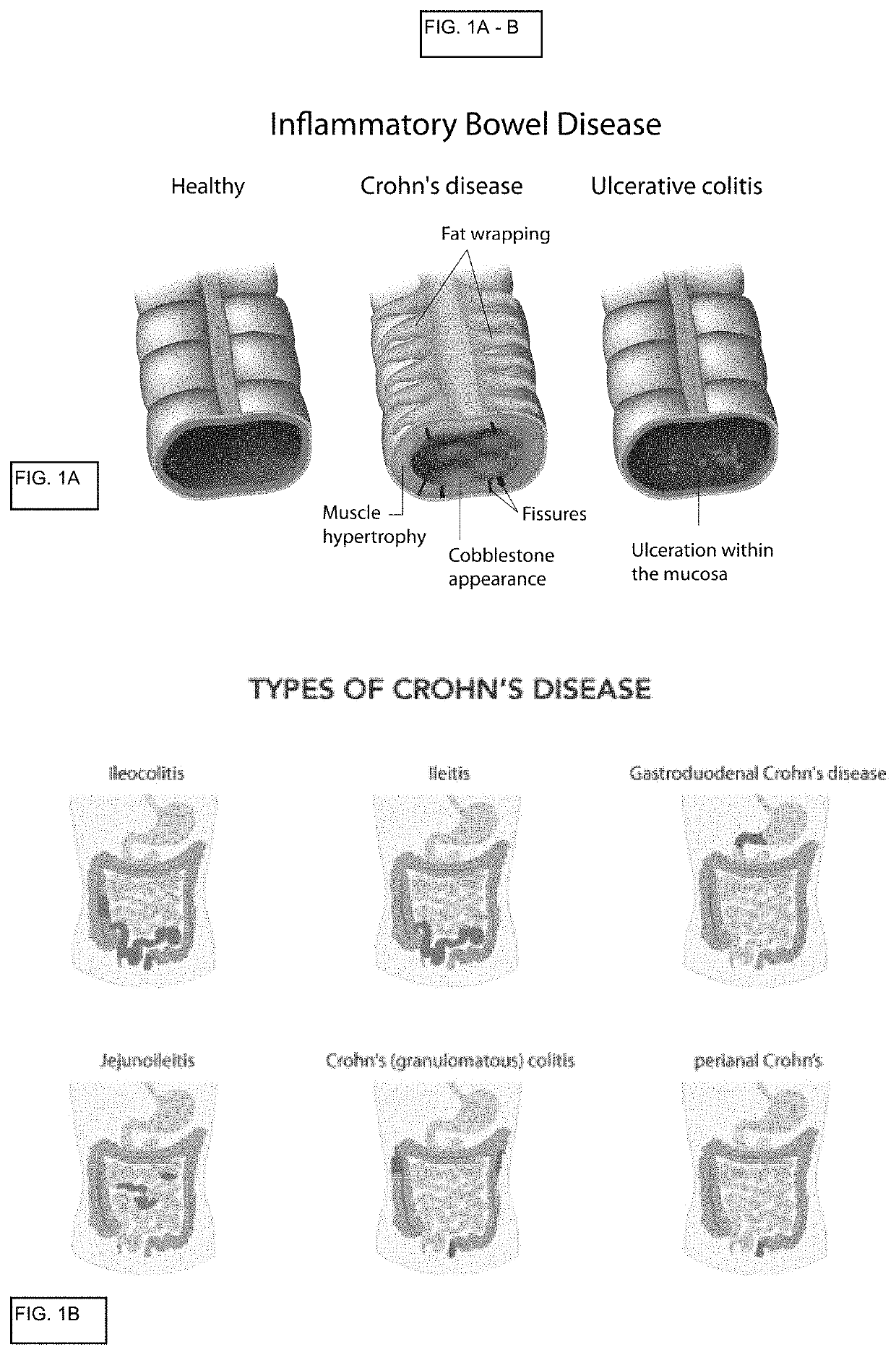
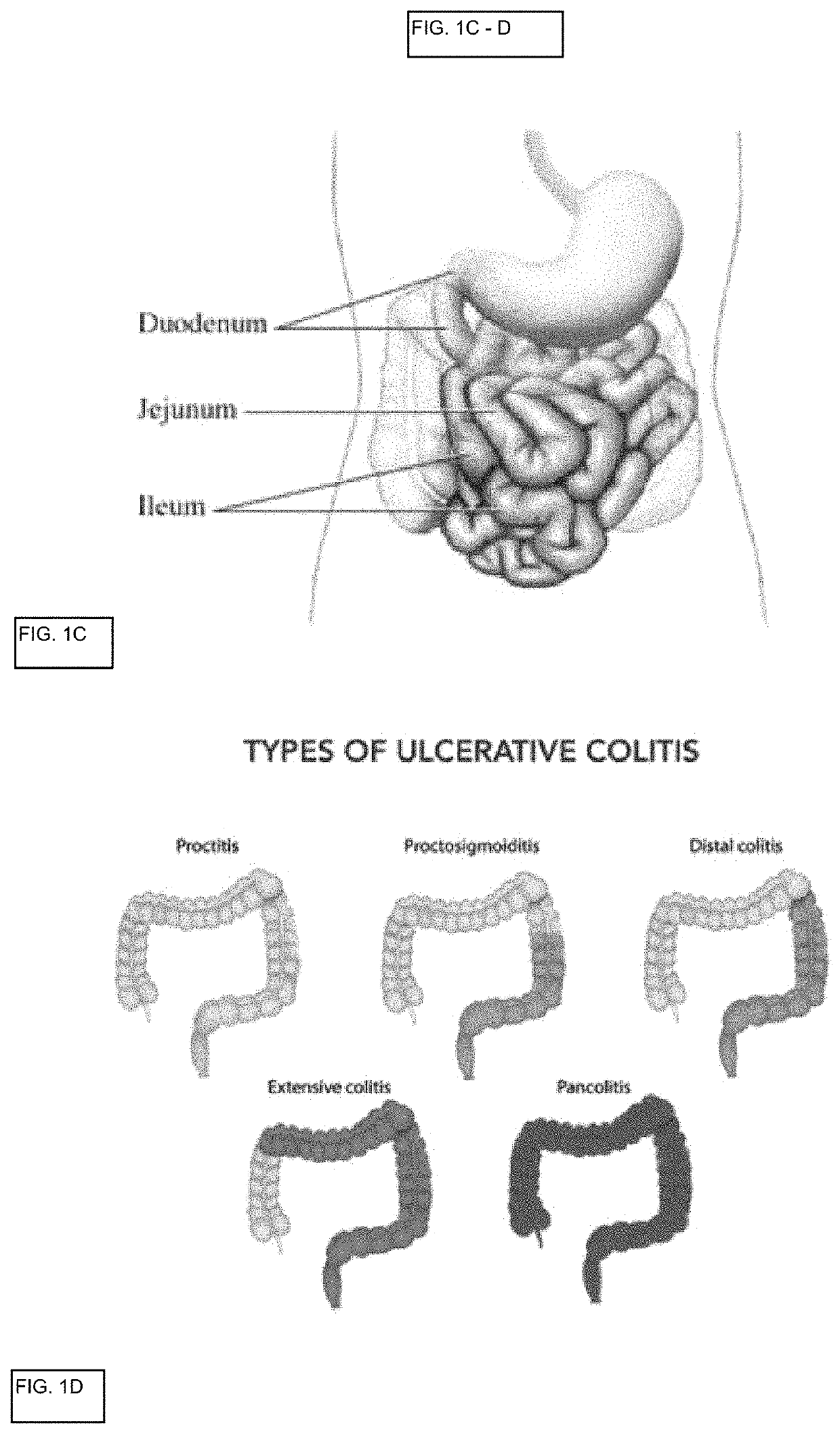
Gut-On-Chip for Modeling Ulcerative Colitis (UC)

[0565]Resident immune cells (B cells, T cells, dendritic cells, macrophages, and innate lymphoid cells) were isolated from healthy and Ulcerative Colitis (UC) patients including inflamed and non-inflamed regions of patient tissue. These cells were used as lamina propria-derived resident immune cells in a Gut-On-Chip as described above.

[0566]A. UC Lamina Propria-Derived Cells Disrupt Epithelial Barrier Function.

[0567]Inflamed UC LP resident immune cells increases permeability of epithelial cells when co-cultured in a device of the present inventions.

[0568]FIG. 10 shows an exemplary disrupted barrier function of around 0.5×10⁻⁷ Papp (cm / s) (apparent permeability) by co-culturing caco-2 epithelial cells and HUVECs with leukocytes isolated from inflamed UC tissue. Untreated controls for comparison use healthy LP derived cells and no LP cells in Gut-On-Chips for comparisons. Treated samples used leukocytes isolated from non-inflamed UC LP c...


PUM


Abstract

An in vitro microfluidic gut-on-chip is described herein that mimics the structure and at least one function of specific areas of the gastrointestinal system in vivo. In particular, a multicellular, layered, microfluidic culture is described, allowing for interactions between lamina propria-derived cells and gastrointestinal epithelial cells and endothelial cells. This in vitro microfluidic system can be used for modeling inflammatory gastrointestinal tissue, e.g., Crohn's disease, colitis and other inflammatory gastrointestinal disorders. These multicellular, layered microfluidic gut-on-chip further allow for comparisons between types of gastrointestinal tissues, e.g., small intestinal deuodejeum, small intestinal ileium, large intestinal colon, etc., and between disease states of gastrointestinal tissue, i.e. healthy, pre-disease and diseased areas. Additionally, these microfluidic gut-on-chips allow identification of cells and cellular derived factors driving disease states and drug testing for reducing inflammation.

Description

FIELD OF THE INVENTION[0001]An in vitro microfluidic gut-on-chip is described herein that mimics the structure and at least one function of specific areas of the gastrointestinal system in vivo. In particular, a multicellular, layered, microfluidic culture is described, allowing for interactions between lamina propria-derived cells and gastrointestinal epithelial cells and endothelial cells. This in vitro microfluidic system can be used for modeling inflammatory gastrointestinal tissue, e.g., Crohn's disease, colitis and other inflammatory gastrointestinal disorders. These multicellular, layered microfluidic gut-on-chip further allow for comparisons between types of gastrointestinal tissues, e.g., small intestinal deuodejeum, small intestinal ileium, large intestinal colon, etc., and between disease states of gastrointestinal tissue, i.e. healthy, pre-disease and diseased areas. Additionally, these microfluidic gut-on-chips allow identification of cells and cellular derived factors ...

Claims


Application Information

Patent Timeline
Patent Type & Authority Patents(United States)
IPC IPC(8): C12M3/06G01N33/50B01L3/00C12N5/077C12N5/071C07K16/28C12M1/12C12N5/0783
CPCC12M23/16B01L3/502753C07K16/2809C07K16/2818C12M25/02C12N5/0636C12N5/0652C12N5/0679C12N5/0697G01N33/505G01N33/5088B01L2300/0681C12N2501/999C12N2502/13C12N2502/23C12N2502/28C12N2503/04C12N2533/90C12N2533/92G01N33/5082G01N2800/065C12M29/10C12M21/08
Inventor KERNS, S. JORDANOBRIGEWITCH, JENIFERSALMON, MICHAELKASENDRA, MAGDALENAUMIKER, BENJAMIN RICHARDS
Owner EMULATE INC
Why Eureka
Try Eureka