Defined three dimensional microenvironment for cell culture

a three-dimensional microenvironment and cell culture technology, applied in cell culture active agents, embryonic cells, peptide sources, etc., can solve the problems of batch-to-batch or lot, experimental variability, inherent heterogeneity, etc., to improve colony shape, increase the surface density of phsrn-rgdsp, and improve the effect of colony siz

Active Publication Date: 2022-01-11
AMOLIFESCI CO LTD +1
View PDF9 Cites 0 Cited by
  • Summary
  • Abstract
  • Description
  • Claims
  • Application Information

AI Technical Summary

Problems solved by technology

These conventional substrates have significant lack of batch-to-batch or lot-to-lot consistency, resulting in experimental variability.
Additionally, current two-dimensional (2D)-based cell culture systems, which suffer from inherent heterogeneity and limited scalability and reproducibility, are emerging as a bottleneck for producing sufficient numbers of high-quality cells for downstream applications.
A major challenge remains with the in vitro expansion and culture of self-renewable stem cells and the subsequent differentiation of these cells.
However, challenges still exist in optimizing the wide variety of platforms capable of supporting cell therapy needs.
However, existing technologies have some limitations to generate a microenvironment that induces a combinatorial signal pathway by selectively, simultaneously or sequentially activating at least two different cell surface receptors in a precisely manner, due to their lack of physical or biochemical attributes.
In addition, various microenvironmental cues are often intertwined and cannot be individually controlled in existing technologies.

Method used


Image

  • Defined three dimensional microenvironment for cell culture
  • Defined three dimensional microenvironment for cell culture
  • Defined three dimensional microenvironment for cell culture

Examples


example 1

Preparation of Electrospinnable Biofunctional Composition to Engineer an Extracellular Microenvironment

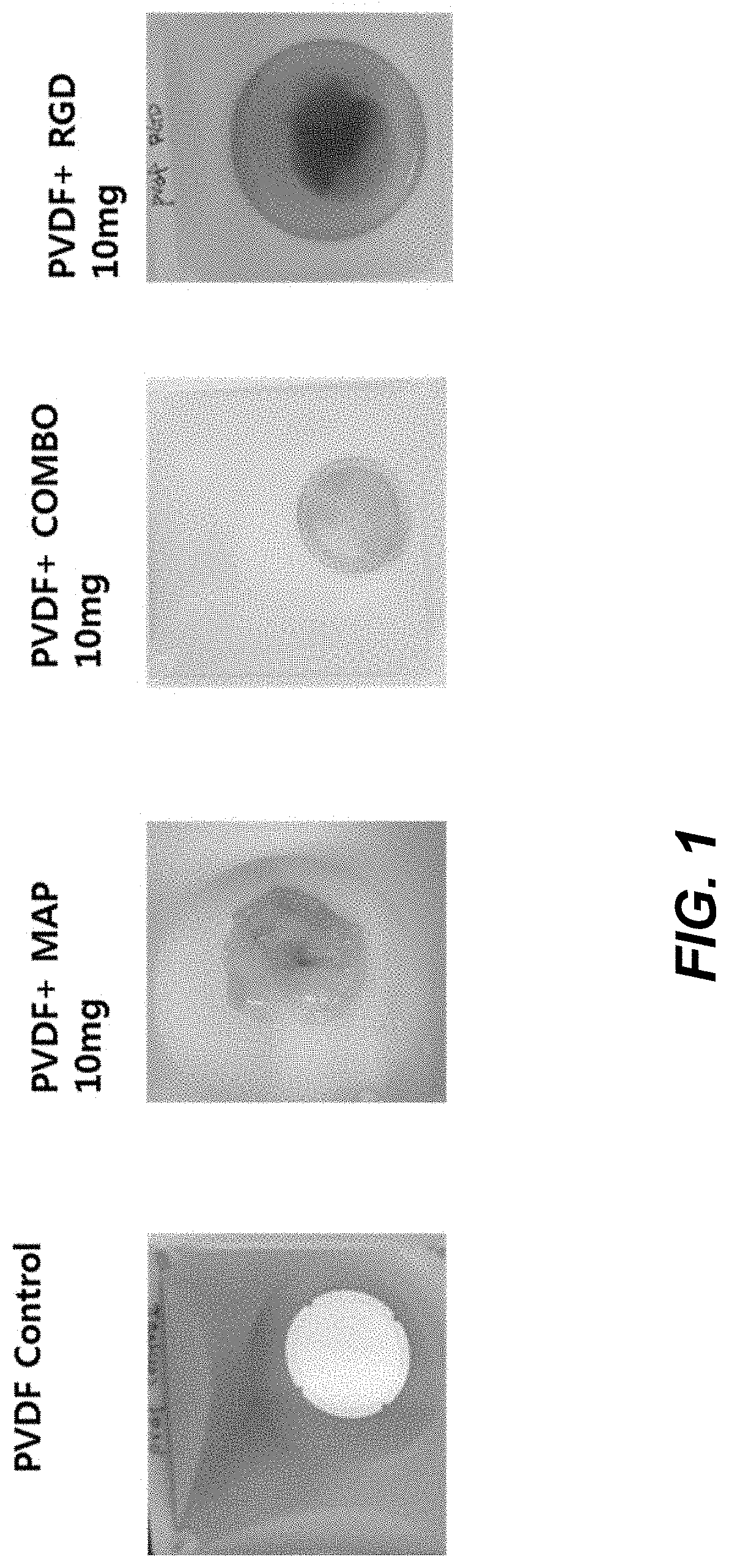
[0128]PVDF with an average molecular weight of 200 kDa from Sigma Aldrich (St. Louis, USA), PAN with an average molecular weight of 200 kDa, PES with an average molecular weight of 200 kDa purchased from Sigma Aldrich (St. Louis, USA), PLA with an average molecular weight of 200 kDa purchased from Sigma Aldrich were dissolved in DMAc to prepare 20 wt % solution. MAPTrix™ ECM (no peptide motif, PHSRN-RGDSP (SEQ ID NO:17) (combo) containing, and RGD motif containing) purchased from Kollodis BioSciences (North Augusta, S.C., USA) was dissolved in an aqueous solution composed of distilled water and DMAc. Each polymer solution was mixed well together with MAPTrix™ ECM solution by vortexing it for 10 minutes to make homogeneous 18 wt % solution.

[0129]The electrospinnable (e-spin) solution was placed in a plastic syringe fitted with a 27 G needle. A syringe pump (KD Scientific, USA) was u...

example 2

Preparation of Nanofiber Having Different Diameter

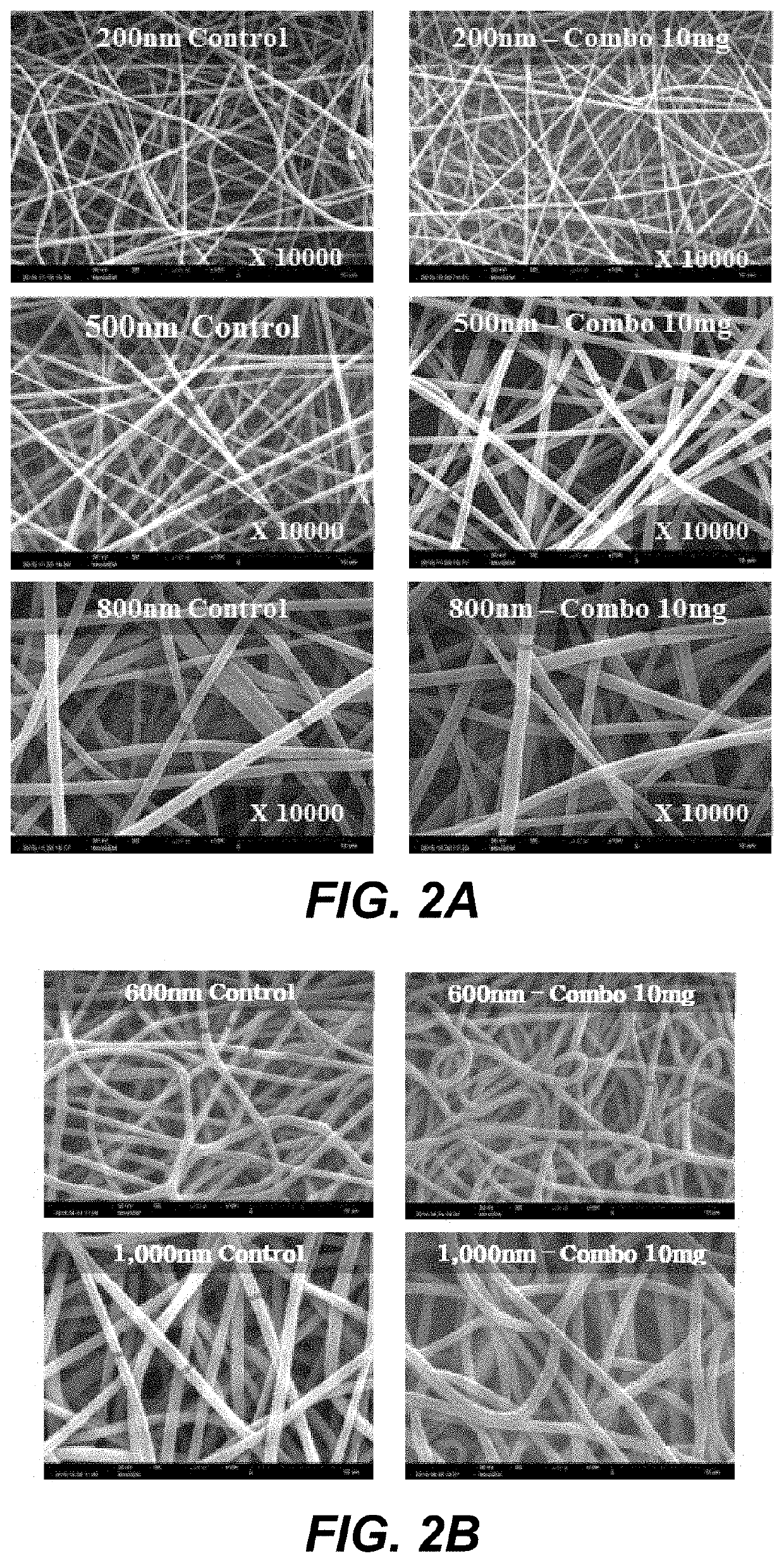
[0136]Each Polyvinylidene fluoride (PVdF)-Kynar 761(Homopolymer, Mw: 400,000-500,000), and Polyvinylidene fluoride (PVdF)-Solef 21216(Co-polymer, Mw: 600,000) or Polyacrylonitril-Pulver (Co-PAN, Mw: 85,000) was dissolved in DMAC and blended. The blending ration of homopolymer to copolymer were 1:1, 1:2, 1:3, 1:4, 1:5, 1:6, 1:7, 1:8, 1:9. Nanofibers having different diameter were formed by the same procedure mentioned in EXAMPLE 1.

[0137]The diameter of each nanofiber sheet is measured by observation using a scanning electron microscope (SEM), a thin gold layer was deposited on the surface of each nanofiber sheet. FIG. 2A shows nanofiber sheets having a diameter of 200, 500, and 800 nm, respectively. FIG. 2B shows nanofiber sheets having a diameter of 600 and 1,000 nm, respectively.

example 3

Preparation of Nanofiber Having Particles

[0138]MAPTrix™ ECM based particles were formed by reaction of the carboxyl group of MAPTrix™ activated by 1-ethyl-3-(3-dimethylaminopropyl) carbodiimides / N-hydroxysulfosuccinimide (EDC / s-NHS) on the C-terminus with the amino groups of the MAPTrix™.

[0139]1-Ethyl-3-[3-Dimethylaminopropyl]carbodiimide hydrochloride (EDC) solution is prepared by dissolving 10 mg of EDC in 1 ml of sodium bicarbonate buffer (10 mM, pH 6.5). 5 mg of solid sulfo-N-hydroxysulfosuccinimide (S—NHS) is added to the EDC solution. The EDC / S—NHS solution is added to the nanofiber surface to activate carboxyl group of MAPTrix™ surfaced on the nanofiber sheet for 30 minutes. After the C-terminus activation, 0.1 mg of MAPTrix™ having PHSRN-RGDSP (SEQ ID NO:17) motif dissolved in 1 mL distilled water was added to the nanofiber surface. Crosslinking is carried out at ambient temperature for 30 minutes to get crosslinked MAPTrix™ particle presenting PHSRN-RGDSP (SEQ ID NO:17) on ...


PUM

PropertyMeasurementUnit
diameteraaaaaaaaaa
diameteraaaaaaaaaa
diameteraaaaaaaaaa

Abstract

Described is a three-dimensional (3D) microenvironment presenting defined biochemical and physical cues that regulate cellular behavior and use of the microenvironment. A composition to form the 3D microenvironment is provided by combining one or more natural or synthetic polymeric materials and substrate proteins recombinantly or chemically functionalized with a variety of bioactive peptides such as extracellular matrix-derived or growth factor-derived peptides. Also described are devices and methods for screening for optimal combinations of the bioactive motifs in order to create an extracellular microenvironment that can regulate specific cellular behavior such as cell growth, proliferation, migration or differentiation.

Description

CROSS-REFERENCE TO RELATED APPLICATION[0001]This application is a continuation-in-part of co-pending U.S. patent application Ser. No. 15 / 173,488, filed Jun. 3, 2016, which itself claims the benefit of the filing date of U.S. Provisional Patent Application Ser. No. 62 / 171,767, filed Jun. 5, 2015, the disclosure of each of which is hereby incorporated herein in its entirety by this reference.TECHNICAL FIELD[0002]This application relates to synthetic microenvironment for culturing cells such as valuable cells including stem cells such as pluripotent stem cells. More particularly, provided are methods and microenvironmental surfaces for culturing pluripotent stem cells, adult stem cells, and adult cells on a defined three-dimensional microenvironment.STATEMENT ACCORDING TO 37 C.F.R. § 1.821(c) or (e)—SEQUENCE LISTING SUBMITTED AS ASCII TEXT FILE[0003]Pursuant to 37 C.F.R. § 1.821(c) or (e), a file containing an ASCII text version of the Sequence Listing has been submitted concomitant wi...

Claims


Application Information

Patent Timeline
Patent Type & Authority Patents(United States)
IPC IPC(8): C12N5/00C12N5/0735
CPCC12N5/0062C12N5/0075C12N5/0606C12N2501/998C12N2513/00C12N2533/30C12N2535/00C12N5/0068C07K14/435
Inventor KIM, CHANBAEK, KYUWONGOO, HUI-GWANLEE, SANGJAEHONG, BONGJINKOO, SONG HEESEO, IN YONGLEE, SEUNG HOONLEE, JI HYUNJANG, SEONHOSEO, DONG-SIK
Owner AMOLIFESCI CO LTD
Why Eureka
Try Eureka