Method for circulating tumor cells isolation

a tumor cell and isolation method technology, applied in tumor/cancer cells, biomass after-treatment, biochemistry apparatus and processes, etc., can solve the problems of cumbersome inspection procedure, inability to achieve standardized testing, and prone to deviation of repeated test results of the same sampl

Active Publication Date: 2022-09-20
NAT TAIWAN UNIV
View PDF1 Cites 0 Cited by
  • Summary
  • Abstract
  • Description
  • Claims
  • Application Information

AI Technical Summary

Benefits of technology

[0025]The present application utilizes the special affinity difference of cellulose and / or its derivatives for different cells to achieve the effect of isolating tumor cells from the blood sample. Cellulose and / or its derivatives themselves have the characteristic that to let the white blood cells in the blood adhere to it. The present application further discloses that the white blood cells are adhered to the cellulose and / or its derivatives but the cancer cells are suspended in the culture medium after a blood sample to be tested culturing on a cell cultural container which its cell adhesion portion is coated with cellulose and / or its derivatives. After the blood containing cancer cells being cultured in the cellulose and / or its derivatives coated-cell cultural container for a period of time, the red blood cells would settle at the bottom, the white blood cells would adhere to the cellulose and / or its derivatives, and the cancer cells would suspend in the culture medium, which achieves the effect of separating cancer cells from the blood. The cancer cells can further be isolated from the cultural system for related analysis and detection to achieve early diagnosis and screening. This isolation process does not use specific biological antibodies. Compare with other products, therefore, the operation is relatively simple, convenient, time-saving and low-cost.

Problems solved by technology

The above methods have their own advantages, but the biggest drawback is the inability to achieve the standardized testing.
Due to various factors, such as reagent composition, operation procedure, and interpretation process, the repeated test results of the same sample are prone to deviation.
The entire detecting process takes about 3-4 hours, so the inspection procedure is cumbersome.
The required specific antibodies are expensive, and the detection cost is relatively high.
In the process of detection, white blood cells and red blood cells in the blood are often prone to false positives in the interpretation results.
Hence, how to separate the blood cells from the cancer cells in the detection system is the biggest challenge.

Method used


Image

  • Method for circulating tumor cells isolation
  • Method for circulating tumor cells isolation
  • Method for circulating tumor cells isolation

Examples


example 1

on of the Isolating Cultural System of Circulating Tumor Cells

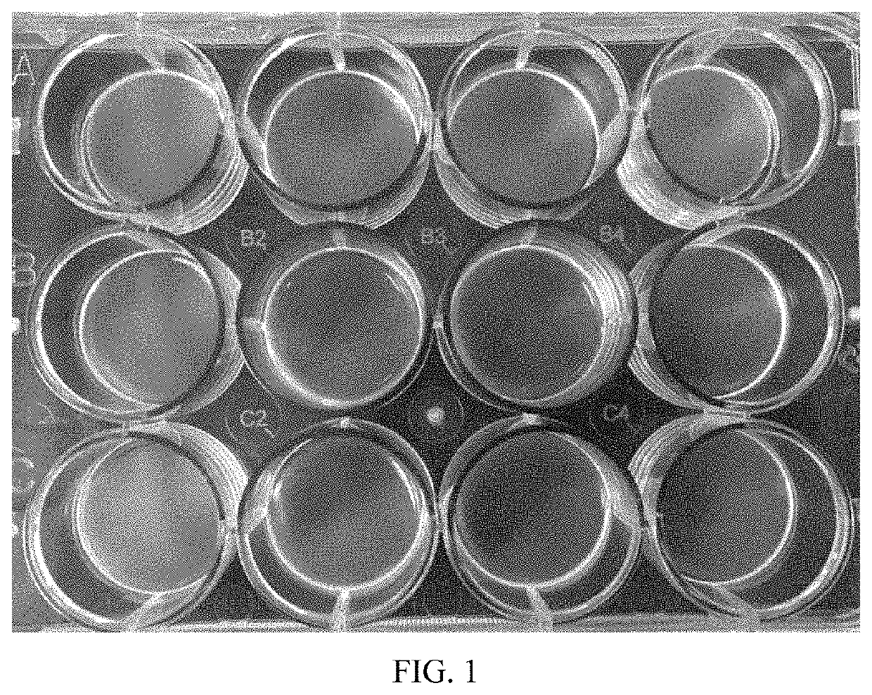
[0063]5 grams of cellulose and / or its derivatives were poured into 100 ml of 5% NaOH. After stirring evenly, the solution is placed at −20° C. for 4 hours to reach complete frozen. Then, the frozen solution was placed at room temperature until completely thawed and was stirred. The freezing-thawing procedures were repeated 5 times until the cellulose solution became translucent. The solution was coated on the culture container in an amount of 12-13 mg / cm2 of cellulose. The coated container was placed at room temperature 26.8-32.6° C., humidity 34.2-52.1%. After drying in the shade for 24 hours, the solid state of the cellulose is gelatinous or jelly-like as shown in FIG. 1. 1 ml of 0.5M HCl was dropped into the coated container and leaving it for 5 minutes. After that, HCl was poured off and the culture container was rinsed twice with filtered water. After sterilization of UV light exposure for 30 minutes, it can be used ...

example 2

od Cells Adhesion Test

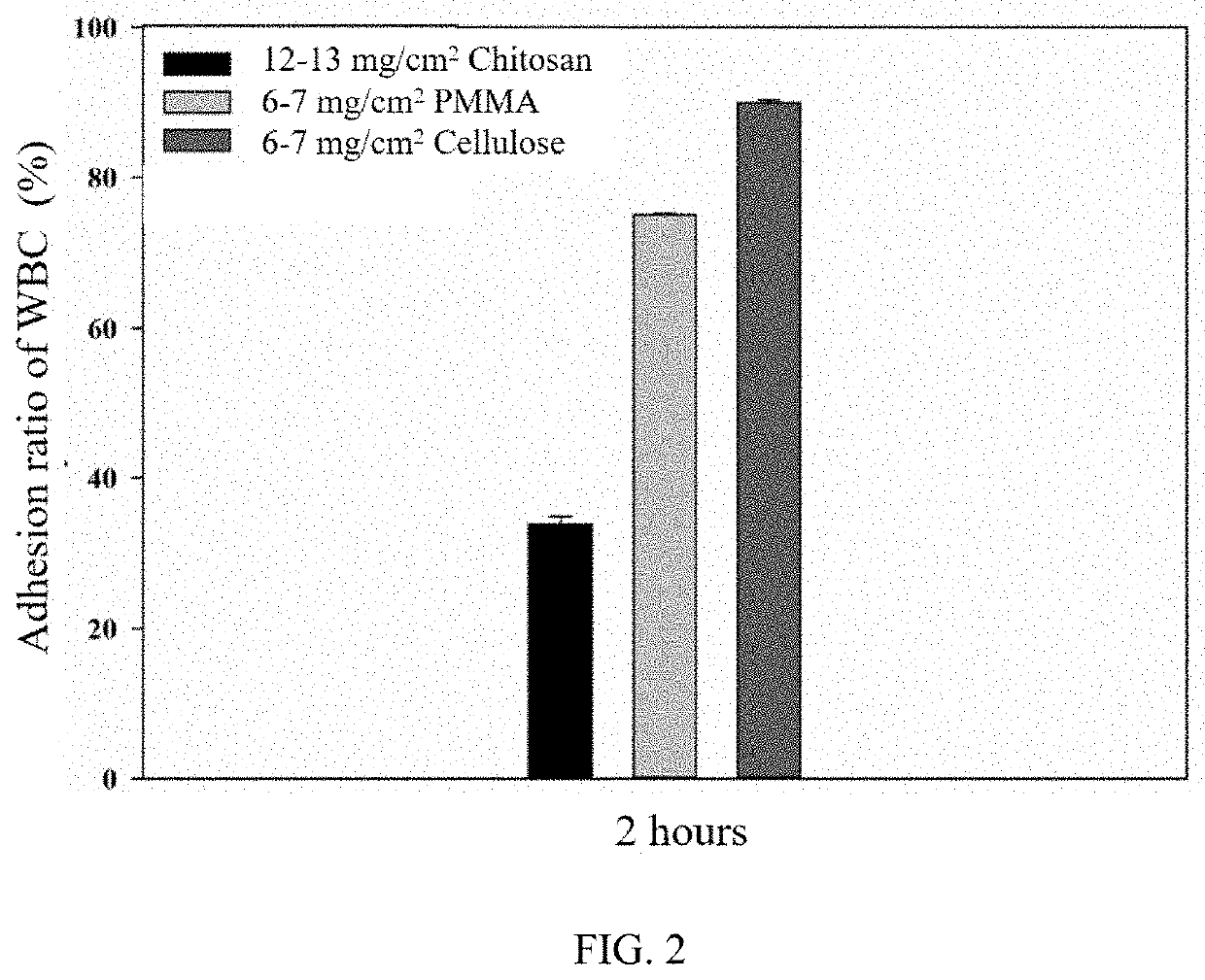
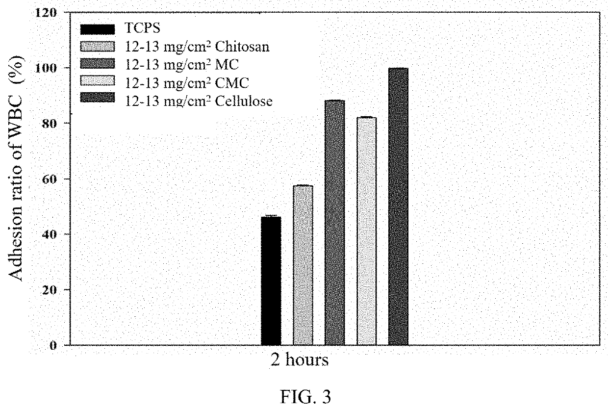
[0065]200 ml of whole blood was taken and was centrifuged at 40 rpm for 20 minutes. The upper layer of the plasma was aspirated off, the buffy coat was obtained, and RBC lysis buffer was added to make the volume ratio 1:3. After uniformly mixing, centrifuge at 15 rpm for 5 minutes and aspirate the supernatant. Saline was added and was mixed well, then centrifuge at 15 rpm for 5 minutes again. The obtained white blood cells were equally divided and cultured in a 10 cm dish, which coated with 12-13 mg / cm2 of Chitosan G214, 6-7 mg / cm2 of PMMA, and 6-7 mg / cm2 of cellulose. The culture dish was placed in 5% CO2 incubator at 37 for 2 hours. After that, supernatant was collected and the number of white blood cells which not adhered to the material was calculated through an automated hematology analyzer. The adhesion ratio of blood cells on the material can be obtained by the following formula:

Adhesion ratio of white blood cells (%)=[(Total number of white blood cells ...

example 3

ls Adhesion Test

[0068]2×10⁴ of fluorescently labeled cells of A549 (human lung adenocarcinoma cells), HT29 (human colon cancer cells) and HeLa (human cervical cancer cells), were separately provided and cultured on TCPS, the culture container coated with 12-13 mg / cm2 of methyl cellulose+cellulose (40:1), the culture container coated with 12-13 mg / cm2 of cellulose for 2 hours.

[0069]FIGS. 4A-4C are the results of A549 human lung adenocarcinoma cells cultured on different materials. FIG. 4A clearly shows that the A549 cells adhere to TCPS. FIG. 4B shows that the A549 cells are suspended in the culture medium within the cell culture container coated with 12-13 mg / cm2 of methyl cellulose+cellulose (40:1). FIG. 4C shows that the A549 cells are also suspended in the culture medium within the cell culture container coated with 12-13 mg / cm2 of cellulose.

[0070]FIGS. 5A-5C are the results of HT29 human colon cancer cells cultured on different materials. FIG. 5A clearly shows that the HT29 cell...


PUM

PropertyMeasurementUnit
temperatureaaaaaaaaaa
adhesionaaaaaaaaaa
cell size isolation methodaaaaaaaaaa

Abstract

A method of circulating tumor cells isolation, using an isolating cultural system of circulating tumor cells, comprises the following steps: (1) providing a sample; (2) adding a cell culture medium to the isolating cultural system of circulating tumor cells; (3) adding the sample to the isolating cultural system of circulating tumor cells to cultivate; and (4) collecting the suspended circulating tumor cells in the cell culture medium; wherein the isolating cultural system of circulating tumor cells comprises a container including a cell adhesion portion, and cellulose coated on the cell adhesion portion.

Description

CROSS-REFERENCE TO RELATED APPLICATIONS[0001]This application claims the priority of Taiwan Application No. 107133389 filed on Sep. 21, 2018 in the State Intellectual Property Office of the R.O.C., the disclosure of which is incorporated herein by reference.BACKGROUND OF THE INVENTIONField of the Invention[0002]The present application relates to a method of circulating tumor cells isolation, and particularly relates to a method for isolating the circulating tumor cells by using a cell cultural container which its cell adhesion portion is coated with cellulose and / or its derivatives.Descriptions of the Related Art[0003]At present, there are many methods for detecting circulating cancer cells (CTCs), including: cell size isolation method, immune magnetic-activated cell sorting method, density gradient centrifuge method. There are also many subsequent analysis methods, including: Immunofluorescence technique, reverse transcription-polymerase chain reaction, flow cytometry, gene chip, e...

Claims


Application Information

Patent Timeline
Patent Type & Authority Patents(United States)
IPC IPC(8): C12N5/09
CPCC12N5/0693C12N5/0694C12N2523/00C12N2533/78C12N5/0093C12M47/04
Inventor YOUNG, TAI-HORNGHUANG, WAN-CHENHSIEH, HAO-YINGLO, SHYH-CHYICHEN, KE-CHENGCHEN, JIN-SHINGCHEN, YIN-TZU
Owner NAT TAIWAN UNIV
Why Eureka
Try Eureka