

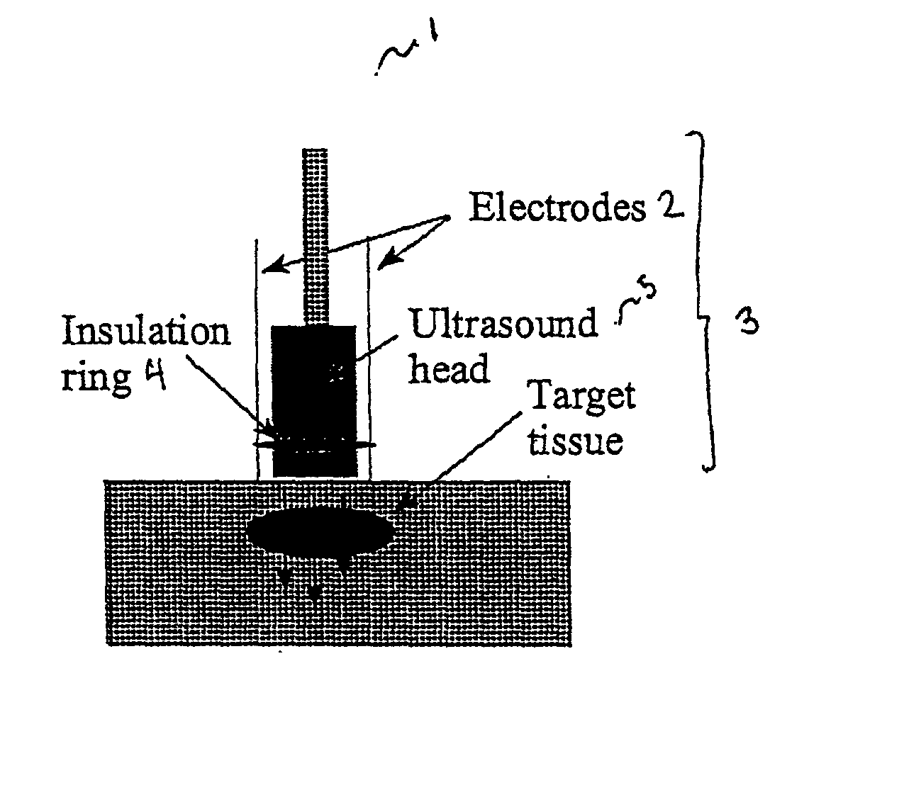
Ultrasound therapy for selective cell ablation
a selective cell and ultrasound technology, applied in the field of ultrasound therapy for selective cell ablation, can solve the problems of low intensity ultrasound rarely causing collateral tissue damage, low intensity ultrasound passing through intervening tissues, and relatively non-destructive
- Summary
- Abstract
- Description
- Claims
- Application Information
AI Technical Summary
Problems solved by technology
Method used
Image

Examples
example 1
The Effect of Low Intensity Ultrasound on Cells Treated with Pulses of 3.625 kV / cm
[0245] The target cell line employed in these studies was a mouse friend leukaemic lymphoblast cell line (clone 707, ECACC no. 91112126 from the European Collection of Animal Cell Cultures) and is maintained in DMEM supplemented with 10% (v / v) foetal bovine serum. Cultures were maintained in a humidified 5% CO.sub.2 atmosphere at 37.degree. C. Cells were harvested by centrifugation, washed once in phosphate buffered saline (PBS) and suspended at a concentration of 1.065.times.10.sup.7 cells / ml. 0.7 ml aliquots of this suspension were dispensed into electroporation cuvettes (0.4 cm electrode gap) together with 0.1 ml of PBS. Cuvettes were retained on ice and electroporated by delivering two pulses of 3.625 kV / cm at a capacitance of 1 .mu.F. Cells were washed twice in PBS by centrifugation, resuspended in PBS containing MgCl.sub.2 (4 mM) (PBS / Mg) and retained at room temperature for 30 minutes. Cells wer...
example 2
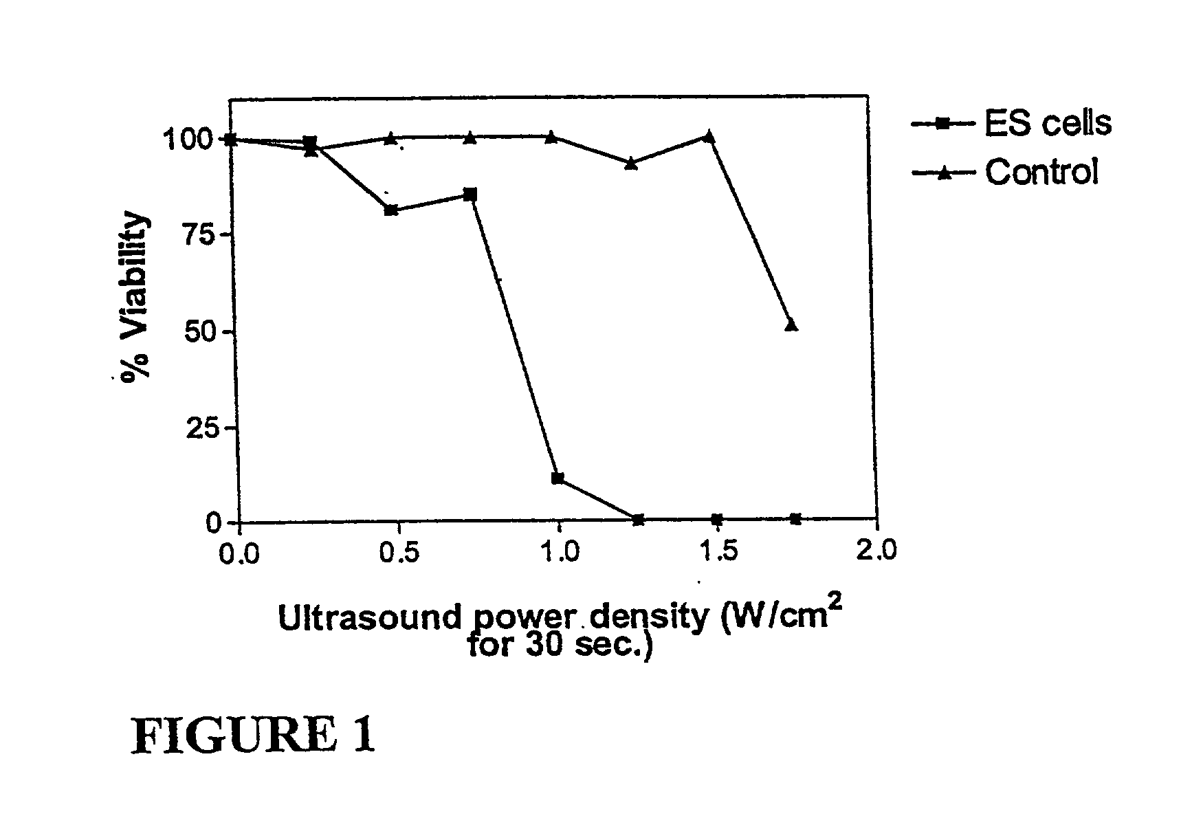
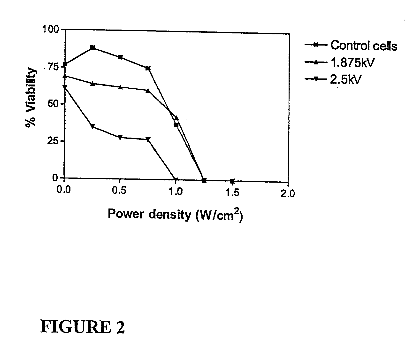
The Effect of Low Intensity Ultrasound on Cells Exposed to Pulses of 1.875 and 2.5 kV / cm
[0247] In order to determine whether or not the pulse electric field strength had any effect on susceptibility of the treated cells to low intensity ultrasound, 0.7 ml aliquots of cells (0.8.times.10.sup.7 cells / ml in PBS / Mg) were dispensed into electroporation cuvettes (0.4 cm electrode gap) together with 0.1 ml of PBS. Cuvettes were retained at room temperature and electroporated as described for Example 1 except that one population was treated with two pulses at 1.875 kV / cm and another was treated with two pulses at 2.5 kV / cm. Cells were transferred to PBS / Mg / glucose and retained at room temperature for 15 minutes. Samples were treated with ultrasound for 30 seconds and allowed to stand at room temperature for 1 hour prior to determining cell viability using trepan blue. A control population of cells was taken through the above treatment except that the electroporation event was omitted.
[0248]...
example 3
Sensitisation and Ultrasound Treatment of Cells Immobilised in Alginate Matrices
[0249] In order to determine whether or not this sensitisation phenomenon could be achieved in a mass of cells, thereby mimicking a tumor mass, it was decided to embed the cells in an alginate matrix, expose the mass to electric pulses and subsequently expose it to ultrasound. Viability could then be determined using a modification of the MTT assay described previously (Rollan et al., Bioprocess Eng. 15: 47, 1996). To the above end 707 cells were harvested and suspended in 1% (w / v) sodium alginate (Keltone LV, Lot no. 35245A, Kelco, UK) at a concentration of 1.16.times.10.sup.7 cells / ml. This suspension were added drop-wise to a calcium chloride solution (1.5% [w / v]) and beads (average vol. per bead=10 .mu.l) retained in CaCl.sub.2 for 15 minutes. Beads were subsequently rinsed in PBS and dispensed into electroporation cuvettes (30 beads / cuvette) together with 0.5 ml PBS. Two electric pulses of 2.5 kV / cm...
PUM
 Abstract
Description
Claims
Application Information