
Method for quantitative determination of glycated hemoglobin

Inactive Publication Date: 2003-04-17
PORTASCIENCE LLC
View PDF0 Cites 34 Cited by
  • Summary
  • Abstract
  • Description
  • Claims
  • Application Information

AI Technical Summary

Problems solved by technology

However, routinely used direct measurement of blood glucose level in patients has a limited value since it gives information only about the glucose concentration at the time of sampling and is influenced dramatically by diet.
a) charge differences as in ion-exchange chromatography [U.S. Pat. No. 4,407,961, U.S. Pat. No. 4,649,122, U.S. Pat. No. 4,270,921, U.S. Pat. No. 4,389,491, U.S. Pat. No. 4,436,820] and in electrophoresis [U.S. Pat. No. 4,351,711]. Ion-exchange chromatography involves separation of hemoglobin fractions in microcolumns of ion exchange resin. Glycated hemoglobin elutes first while the non-glycated hemoglobin remains attached to the resin and can be removed by changing the eluting buffer. For proper separation, the composition, pH and ionic strength of the eluting buffer must be maintained within narrow limits. Additionally, the temperature control is critical. Subsequent spectrophotometrical measurement provides the amount of the fraction constituting the glycated hemoglobin. Electrophoresis exploits relative mobility of the hemoglobin fractions in a specially prepared agar medium in an electric field. Common drawbacks for these methods are poor reproducibility, sensitivity to variations in temperature, pH, ionic strength, and sample storage conditions. Also they require expensive equipment and usually prove too slow and cumbersome for practical use.
b) structural characteristics of the carbohydrate groups on the hemoglobin, as in immunoassay [U.S. Pat. No. 4,247,533,U.S. Pat. No. 4,970,171,U.S. Pat. No. 5,206,144]and boronate affinity chromatography [U.S. Pat. No. 4,269,605]. The major limitation of the immunoassay approach is the requirement of an additional independent measurement of the total hemoglobin in order to express the glycated hemoglobin level as a percentage number. Additionally, both immunoassay and high performance chromatography require very expensive reagents and equipment.
As it was mentioned above, the need of using antibody makes the method rather expensive.
However this technique suffers from rather complex procedure which requires several manual operations.

Method used


Image

  • Method for quantitative determination of glycated hemoglobin
  • Method for quantitative determination of glycated hemoglobin
  • Method for quantitative determination of glycated hemoglobin

Examples


example 1

Manual Format

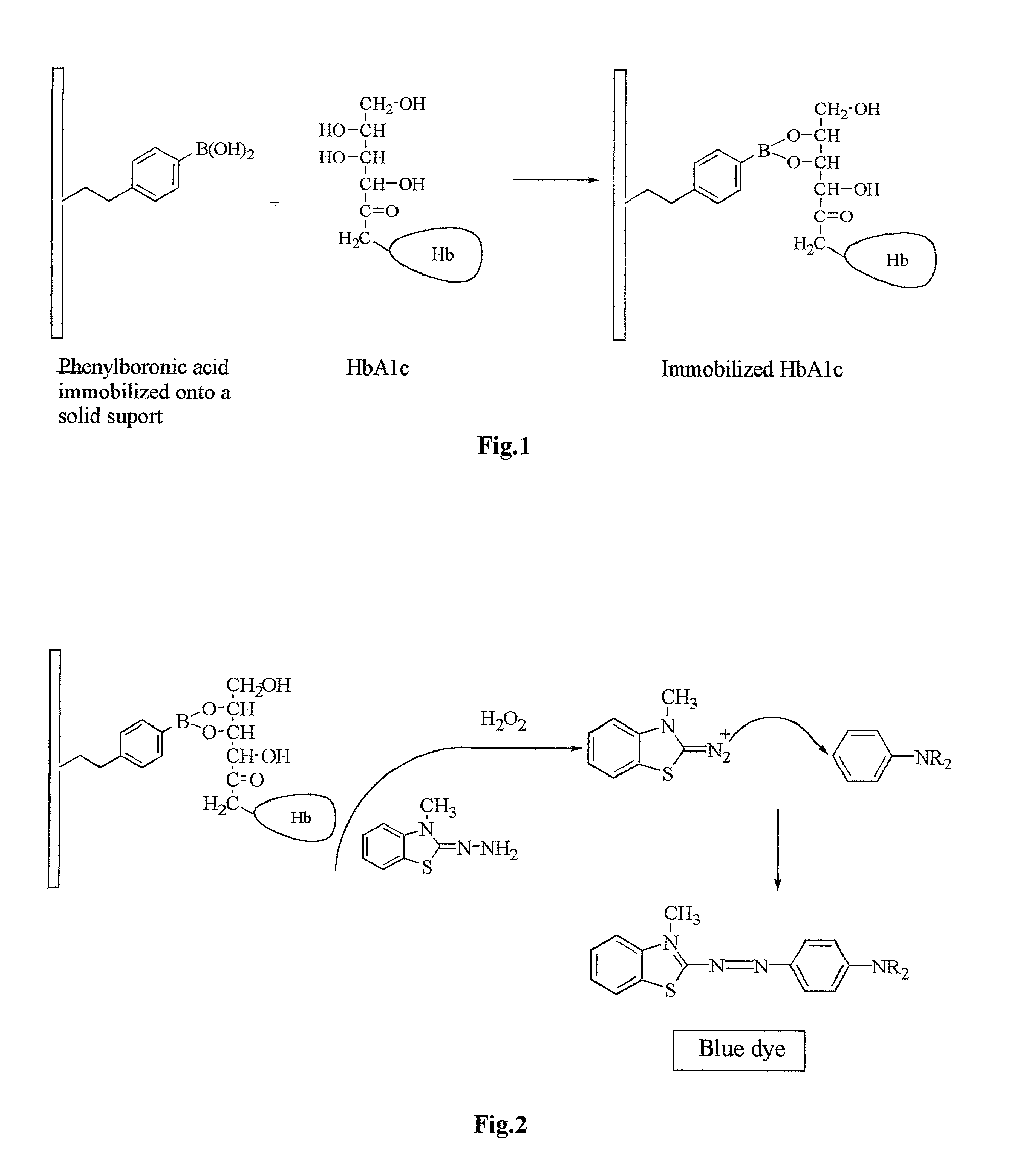
[0035] Whole blood sample (100 .mu.l) was mixed with 250 .mu.l of binding buffer (Taurine, pH 9) and the mixture was incubated with 25 mg of 3-aminophenylboronic acid immobilized onto acrylic beads (Sigma, Cat.# A 4046, Ligand immobilized: 300-600 .mu.moles per gram)) for 10 minutes. The beads were filtered off and washed with phosphate buffer (50 mM, pH 7). The beads were transferred into a vial, and color development solutions containing the color forming coupler and the oxidizable color-developing compound were added in the following order:

[0036] phosphate buffer (50 mM, pH 7) . . . 2.7 ml

[0037] 3-methyl-2-benzothiazolinone hydrazone hydrochloride solution (MBTH)(0.035%) . . . 100 .mu.l

[0038] N,N-diethylaniline (0.08M) . . . 100 .mu.l

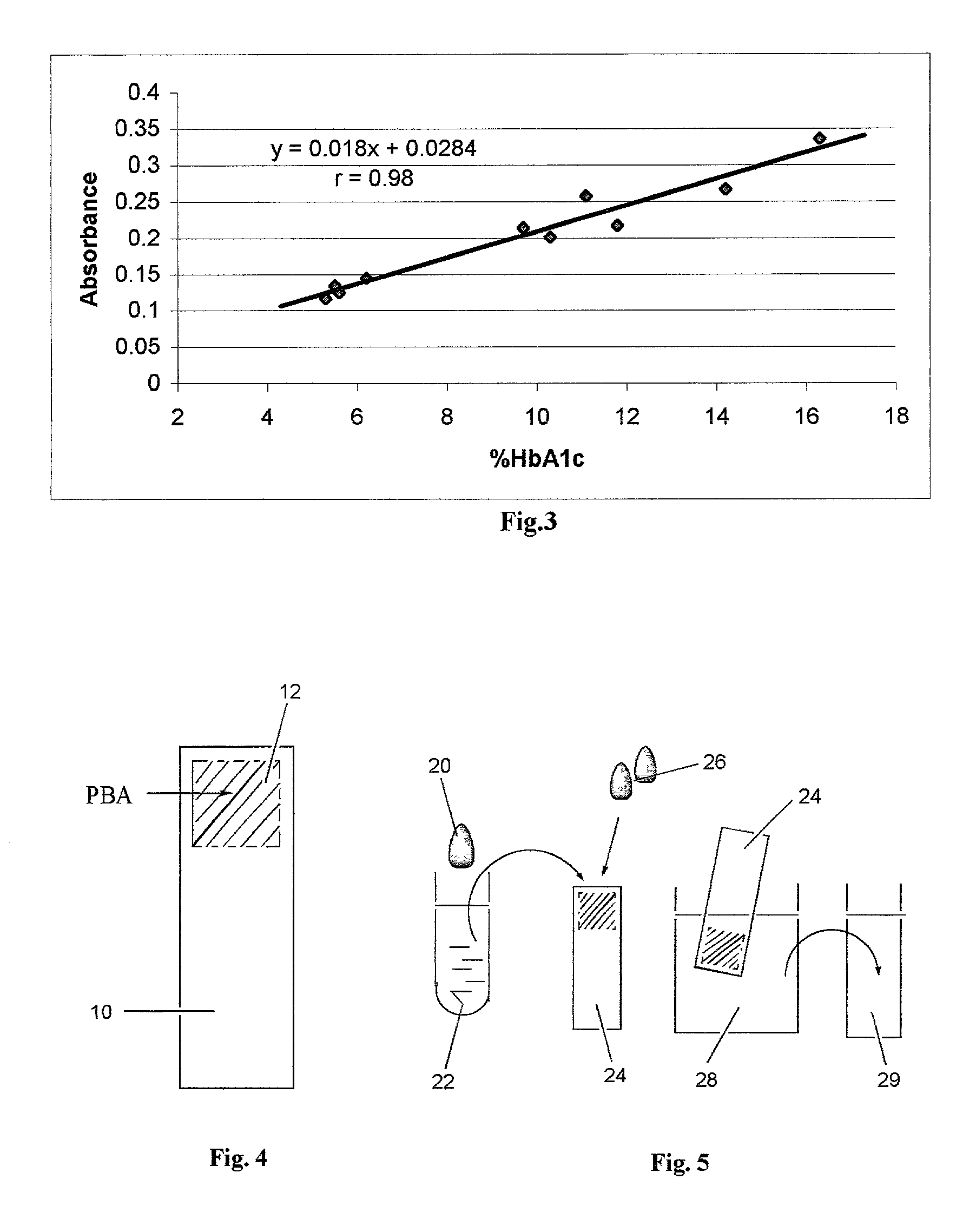
[0039] Finally 100 .mu.l of H.sub.2O.sub.2 (20 mM) was added. The mixture was gently agitated at room temperature for 3 minutes, and the absorbance readings at 596 nm were measured on a spectrophotometer. Ten blood samples with HbA.sub.1...

example 2

Dip Stick Format (Spectrophotometric Measurement)

[0041] The key component of the proposed device is a membrane attached to a plastic strip (FIG. 4). The membrane 12 contains immobilized phenylboronic acid (PBA) and is attached to a backing 10. Biodyne.RTM. C membrane (Pall Gelman Lab., Ann Arbor, Mich.) used in this example is a nylon 6,6 membrane with pore surface populated by carboxylic groups. Onto this membrane is immobilized 3-Aminophenylboronic acid (APBA) using dicyclohexylcarbodiimide (DCC) to form covalent amide bonds.

[0042] Immobilization was accomplished by incubating of the membrane with a solution of APBA and DCC in tetrahydrofuran (THF) for two days at ambient temperature. An excess of APBA was used to block all carboxylic groups and prevent nonspecific binding. In order to remove reaction components, the membrane was washed successively with dimethylformamid (DMF), THF and acetone. The washing continued until the washing solution showed no absorbance in the 200-300 nm...

example 3

Dip Stick Format (Reflectometric Measurement)

[0065] In order to fix dye formed during the test to the membrane surface, NEAP was immobilized onto the membrane along with APBA. A piece of Biodyne C Membrane (4.times.4 cm) was incubated with a solution of APBA (80 mg) and DCC (0.2 g) in dry THF (8 ml) for 24 hours. The membrane was washed by agitating with following solvents for 15 min each: THF (4.times.15 ml), DMF (15 ml), THF (15 ml), and acetone (2.times.15 ml). The washed membrane was briefly dried under the hood and incubated for 24 hours with a solution of NEAP 2HCl (0.25g), diisopropylethylamine (0.4 ml) and DCC (0.8g) in dry DMF (10 ml). The membrane was washed by agitating with following solvents for 15 min each: THF (2.times.15 ml), DMF (15 ml), THF (15 ml), and acetone (2.times.15 ml). A dip stick was produced from the dried membrane as in Example 2.

[0066] The assay procedure was generally the same as in Example 2 except that the colored dye is trapped / fixed on the membran...


PUM


Abstract

A method and a system for separating glycated hemoglobin from non-glycated hemoglobin, and a method for one-read quantitative determination of the glycated hemoglobin and %HbA.sub.1c that does not require an additional measurement of total hemoglobin.

Description

FIELD OF THE INVENTION[0001] The present invention relates to a novel method for measuring the concentration of glycated hemoglobin (gHb) and the percent hemoglobin A.sub.1c (HbA.sub.1c) in blood without using antibodies, and more particularly, to a one-read method which does not require an additional measurement of total hemoglobin for assessing percentage hemoglobin A.sub.1c.BACKGROUND OF THE INVENTION[0002] The importance of diagnosis and monitoring of diabetes is emphasized by the American Diabetes Association reporting that 15.7 million Americans have diabetes, or 5.9% of the population. However, routinely used direct measurement of blood glucose level in patients has a limited value since it gives information only about the glucose concentration at the time of sampling and is influenced dramatically by diet. Nevertheless, an accurate index of a person's mean blood glucose level over 2 to 3 months can be provided by measurement of a specific type of glycated hemoglobin called t...

Claims


Application Information

Patent Timeline
IPC IPC(8): G01N33/72
CPCG01N33/723
Inventor LAW, WAI TAKNIKOLYUKIN, YURI
Owner PORTASCIENCE LLC
Learn More