Novel therapeutic vaccine formulations

a vaccine formulation and technology of a new type of vaccine, applied in the direction of biocide, plant growth regulator, cancer antigen ingredient, etc., can solve the problems of monoclonal antibody, less efficient treatment, and many serious problems

Inactive Publication Date: 2004-02-26
PHARMEXA
View PDF0 Cites 22 Cited by
  • Summary
  • Abstract
  • Description
  • Claims
  • Application Information

AI Technical Summary

Benefits of technology

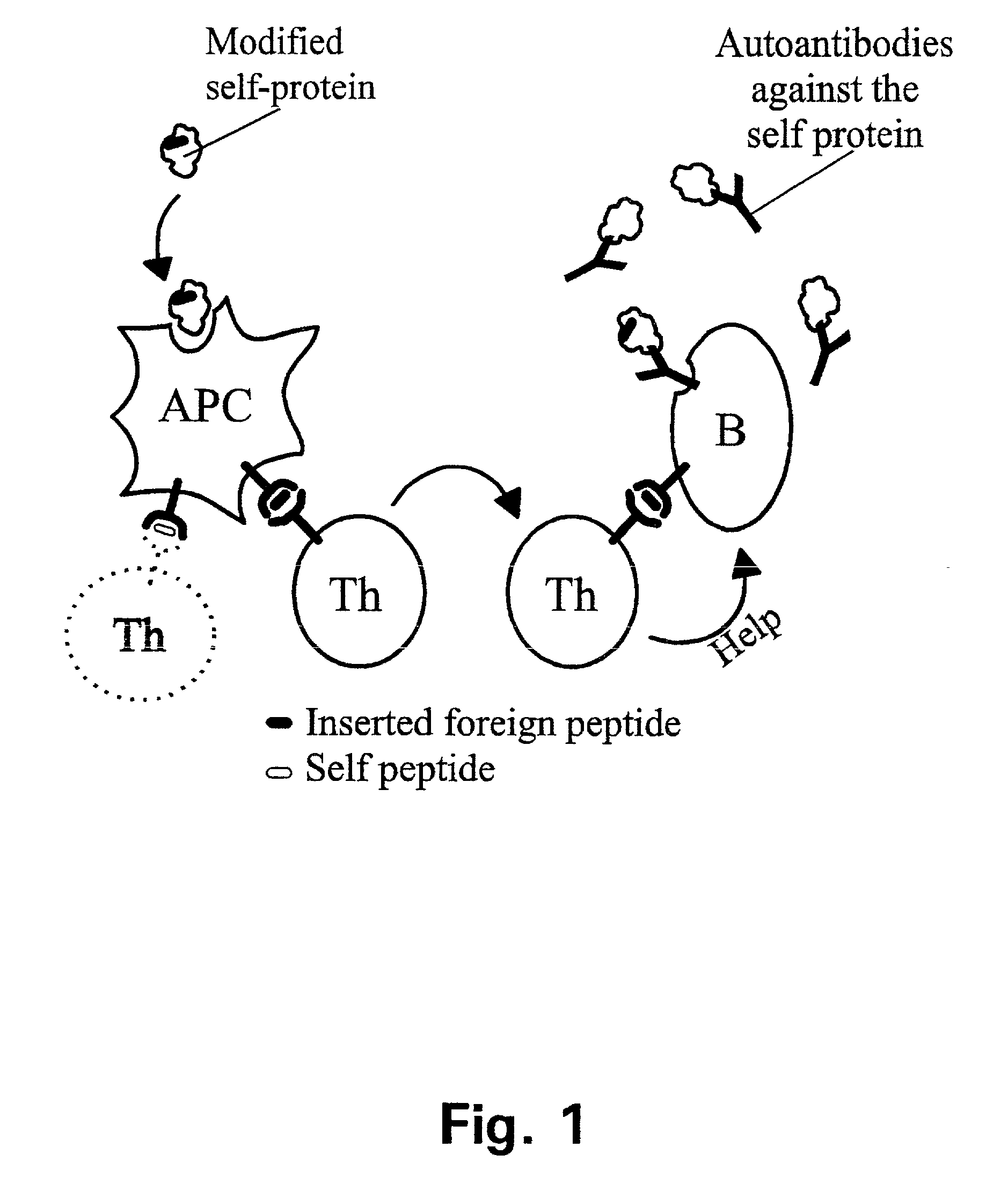
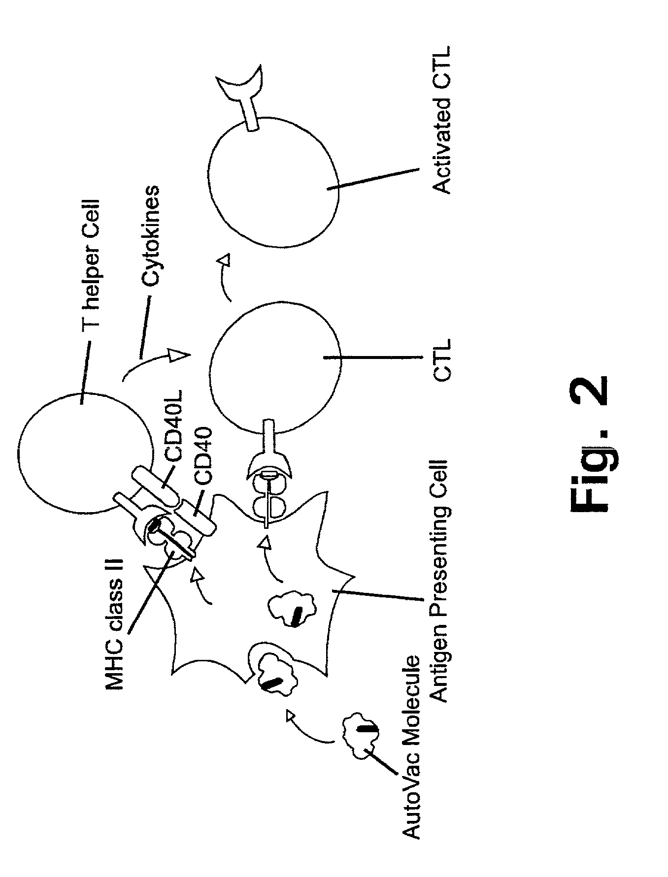
0034] The autovaccine technology described in WO 95 / 05849 has the effect that specific T cell help is provided to self-reactive B cells when a modified self-antigen is administered for uptake into the MHC class II antigen processing pathway (cf. FIG. 1, and Dalum I et al., 1996, J. Immunol. 157: 4796-4804 as well as Dalum I et al., 1999, Nature Biotechnol. 17: 666-669). It was shown that potentially self-reactive B-lymphocytes recognizing self-proteins are physiologically present in normal individuals. However, in order for these B-lymphocytes to be induced to actually produce antibodies reactive with the relevant self-proteins, assistance is needed from cytokine producing T-helper lymphocytes (T.sub.H-cells or T.sub.H-lymphocytes) Normally this help is not provided because T-lymphocytes in general do not recognize T-cell epitopes derived from selfproteins when presented by antigen presenting cells (APCs). However, by providing an element of "foreignness" in a self-protein (i.e. by introducing an immunologically significant modification), T-cells recognizing the foreign element are activated upon recognizing the foreign epitope on an APC (such as, initially, a mononuclear cell). Polyclonal B-lymphocytes (which present T-cell epitopes) capable of recognising self-epitopes on the modified self-protein also internalise the antigen and subsequently presents the foreign T-cell epitope(s) thereof, and the activated T-lymphocytes subsequently provide cytokine help to these self-reactive polyclonal B-lymphocytes. Since the antibodies produced by these polyclonal B-lymphocytes are reactive with different epitopes on the modified polypeptide, including those which are also present in the native polypeptide, an antibody cross-reactive with the non-modified self-protein is induced. In conclusion, the T-lymphocytes can be led to act as if the population of polyclonal B-lymphocytes have recognised an entirely foreign antigen, whereas in fact only the inserted epitope(s) is / are foreign to the host. In this way, antibodies capable of cross-reacting with non-modified self-antigens are induced.

Problems solved by technology

Monoclonal antibody therapy, however, gives rise to several serious problems:
Injection of these foreign substances induces an immune response in the patient towards the injected antibodies, which may lead to less efficient treatment as well as to serious allergic side-effects in the patients.
This is a problem, since the production costs of monoclonal antibodies are huge.
Monoclonal antibodies are usually not able to activate secondary effector systems of the immune system such as complement, NK-cells or macrophage killing of tumour cells.
The latter disadvantage is of particular importance in cancer therapy and may be an important reason why monoclonal antibody therapy of cancer in several cases has not been particularly successful.
The so-called humanised monoclonal antibodies now used by many companies are less immunogenic, but unfortunately they are even less capable of activating the secondary immune effector systems.
Furthermore, examples of secondary outgrowth of tumours lacking the original tumour antigen have been observed, since these antibodies do not induce "innocent bystander" effects on tumour cells not carrying the tumour antigen.
The poor effector capability of the monoclonal antibodies has led to the development of monoclonal antibodies chemically conjugated to different toxins and radioisotopes.
Both constructs are more effective than the monoclonal antibody alone, but they are also more expensive and immunogenic.
Antibodies conjugated to radioisotopes are also expensive as well as immunogenic and other general toxic side-effects are observed.
However, being monoclonal these antibodies only react with a single type of epitope on a tumour antigen.
However, such antibodies induce a vigorous immune response towards the injected foreign polyclonal antibodies which rapidly eliminate the therapeutic effects.
These small carbohydrate structures are, however, very poor antigens so conjugates of these molecules with carrier molecules such as keyhole limpet haemocyanin (KLH) or sheep mucins (containing Tn- and sTn) must be used.
Another example of the active induction of polyclonal antibodies in cancer is the use of idiotype specific vaccination against B-cell lymphomas, which--although it has been promising--is limited to this cancer type only.
However, these cells are somehow rendered non-responsive or anergic by several different possible mechanisms including secretion of immunosuppressive cytokines by the tumour cells, lack of co-stimulatory signals, down regulation of MHC class I molecules etc.
Such peptides have been used to induce a tumour specific immune response in the host, but the practical use of tumour specific peptides in vaccines is restricted to a limited segment of the population due to the narrow HLA class I binding specificity of the peptides.
Furthermore, it is usually relatively difficult to evoke a CTL response in vivo using synthetic peptides due to the low biological half-life of these substances as well as the difficulties with exogenous priming of MHC class I molecules.
Apart from the fact that these treatments usually are very expensive and difficult to reproduce, it has also turned out to be difficult to obtain a good immune response towards the tumour since many of the tumour associated antigens are true self-proteins to which most T cells appear to be tolerant.

Method used


Image

  • Novel therapeutic vaccine formulations
  • Novel therapeutic vaccine formulations
  • Novel therapeutic vaccine formulations

Examples


example 1

[0190] Preparation of Chitosan Microparticles

[0191] 0.2 g of chitosan base (ChitoClear.TM. 804 from Primex Ingredients, Viscosity: 12 mPas measured in 1% in 1% acetic acid, deacetylation: 98.3%) and 0.8 g of Tween 80 is weighed out in a 100 ml beaker and brought into solution by addition of 80 ml of 2% acetic acid and subsequent stirring so as to obtain a solution of 0.25% chitosan, 1% Tween 80 and 2% acetic acid.

[0192] The beaker is placed in an ultrasound probe device (Soniprep 150, MSE) with a magnet stirring device. The solution is sonicated with a small probe for 30 min at 6 mA and magnetic stirring. Initially, sodium sulphate solution is added dropwise until particles precipitate (the amount and concentration can vary, e.g. 2 ml 10% sodium sulphate, 1 ml 20% sulphate etc.).

[0193] The particles are spun down in two 50 ml tubes at 5000 rpm for 20 min (Stratos Biofuge, Heraeus Instruments). The supernatant is isolated and resuspended in MilliQ water. Each batch is pooled in a tub...

example 2

[0197] Loading of Chitosan Microparticles with Ovalbumin

[0198] Solutions of 20 mg / ml chitosan particles in water are prepared as well as solutions of 20 mg / ml ovalbumin in water. 0.5 ml of each solution are mixed in an Eppendorf tube which is left to incubate for 3 hours at room temperature.

[0199] After 3 hours, the suspension is transferred to a 10 ml tube and 4 ml MilliQ is added. The resulting mixture is centrifuged at 10,000 rpm for 15 min. The supernatant is removed by suction and the pellet is resuspended in 5 ml MilliQ water. The mixture is centrifuged again. This procedure is repeated 3 times. The amount of ovalbumin in the supernatant (i.e. non-bound ovalbumin) is determined by means of a BCA assay:

[0200] A standard solution of ovalbumin in water containing 0.5 mg / ml is prepared. This standard is diluted to 0.4, 0.2, 0.1, 0.05 and 0.0125 mg / ml. 20 .mu.l per well of each of these 7 standards as well as a blind are added in triplicate to a flat-bottomed microtiter plate, cf. ...

example 3

[0203] Assaying of CTL Induction and T-Cell Proliferation

[0204] Mice have been injected subcutaneously with the following:

[0205] 1. 200 .mu.l ovalbumin-loaded (0.5 .mu.g / ml) chitosan particles prepared as above (10 .mu.g chitosan per 5 .mu.g ovalbumin).

[0206] 2. 200 .mu.l ovalbumin / chitosan mixture (0.5 .mu.g / ml ovalbumin and approximately same ratio between chitosan and ovalbumin).

[0207] 3. 200 .mu.l ovalbumin in Freund's complete adjuvant (0.5 .mu.g / ml ovalbumin).

[0208] 4. 200 ul of the peptide SIINFEKL (a known CTL epitope from ovalbumin) in Freund's complete adjuvant (0.5 .mu.g / ml SIINFEKL).

[0209] 5. 200 .mu.l ovalbumin in H.sub.2O (0.5 .mu.g / ml ovalbumin).

[0210] 6. 200 .mu.l H.sub.2O.

[0211] Ten days after last immunization, the mice were sacrificed and axillar and inguinal lymph nodes and the spleens were excised.

[0212] In a standard Chrome release assay for determination of CTLs lysing SIINFEKL-carrying cells, results have been obtained showing that CTLs were induced by the al...


PUM

PropertyMeasurementUnit
mean diameteraaaaaaaaaa

Abstract

The present invention relates to a novel method and formulation for the induction of immune responses against polypeptide antigens. In particular, the invention provides a method and formulation for induction of cytotoxic T cell responses against a polypeptide antigen of choice. The formulations are characterized by containing chitosan in admixture with the polypeptide antigen, preferably in the form of microparticles that may be cross-linked.

Description

[0001] The present invention relates to novel methods for combatting diseases, such as cancers, which are characterized by the presence of gene expression products which are non-immunogenic or poorly immunogenic. In particular, the present invention relates to methods for inducing an immune response conducted by cytotoxic T-lymphocytes (CTLs), whereby cells carrying epitopes from the gene expression products are attacked and killed by the CTLs. The invention specifically relates to formulation in chitosan and other chitin-derivatives of selfproteins and "immunogenized" self-proteins in order to provide for enhanced specific immune responses, especially enhanced CTL responses.[0002] Hence, the invention relates to a series of applications of vaccination technology, e.g. within the field of therapeutic vaccination against cancer, but also within the general field of protein vaccination where CTL responses are desired.[0003] The idea of vaccinating against cancer has been around for mo...

Claims


Application Information

Patent Timeline
Patent Type & Authority Applications(United States)
IPC IPC(8): A61K39/00A61K39/39
CPCA61K39/0011A61K39/39A61K2039/57A61K2039/55583A61K2039/5154A61K39/001129A61K39/001112A61K39/001113A61K39/001128A61K39/001176A61K39/001194A61K39/00117A61K39/001191A61K39/001124A61K39/001133A61K39/001157A61K39/001164A61K39/001182A61K39/001197A61K39/001156A61K39/001189A61K39/001192A61K39/001104A61K39/001152A61K39/001103A61K39/001184A61K39/001119A61K39/001188A61K39/001106A61K39/001151A61K39/001172A61K39/001134A61K39/001135A61K39/001144A61K39/001159A61K39/001168A61K39/001171A61K39/001186Y02A50/30
Inventor BEIER, ANNE METTEGAUTAM, ANANDMOURITSEN, SOREN
Owner PHARMEXA
Why Eureka
Try Eureka