Fluorescent proteins from aquatic species

a technology of fluorescent proteins and aquatic species, applied in the field of fluorescent proteins from aquatic species, can solve the problems of high gfp expression, affecting the utility of a protein in research, and toxic to some mammalian cells accumulated protein,

Active Publication Date: 2004-06-10
MIAMI UNIVERISTY OF
View PDF35 Cites 9 Cited by
  • Summary
  • Abstract
  • Description
  • Claims
  • Application Information

AI Technical Summary

Problems solved by technology

However, factors other than fluorescence color and intensity affect the utility of a protein in research.
The stability of many fluorescent proteins makes them undesirable reporters to use if one seeks to determine short term or repetitive events.
Moreover, accumulated protein can be toxic to some mammalian cells.
Although the inventors do not wish to be limited to a single explanation of the toxicity of GFP from Aequorea, it is believed that this is probably due to free radical (H.sub.2O.sub.2) formation which occurs in a 1:1 stoichiometry with GFP production, making high levels of GFP expression particularly toxic.
This results in loss of fluorescence of the fluorophore.
This process is usually reversible but can limit the usefulness of GFP expression, e.g. by reducing time available to photograph specimens.

Method used


Image

  • Fluorescent proteins from aquatic species
  • Fluorescent proteins from aquatic species
  • Fluorescent proteins from aquatic species

Examples


example 1

Species Collection and Animal Husbandry

[0070] Coral colonies were obtained from two sources. First, a solid, brick-red colony consisting of several Actinodiscus / Discosoma sp. polyps was obtained from a local aquarium store. Second, Montastraea cavernosa colonies were collected from reefs in the South Florida area. Both species were maintained in small aquariums with flow-through, filtered sea water.

example 2

Isolation of Total RNA, Reverse Transcription, Amplification and Recovery of cDNAs

[0071] RNA was isolated using the Totally RNA kit (Catalog # 1902, Ambion, Inc., Austin, Tex.) from a single polyp (Actinodiscus / Discosoma sp.) or from a cellular mass lightly airbrushed from the underlying skeleton (M. cavernosa). Briefly, the tissue was homogenized and mixed by tube inversion with approximately 10 volumes of denaturation solution and extracted with 1 volume of phenol / chloroform. The aqueous supernatant was transferred to a clean vessel. One-tenth volume of 3M sodium acetate was added to the supernatant, which was then extracted an additional time with 1 volume of acid-phenol / chloroform. Again, the aqueous supernatant was transferred to a clean vessel. One volume of isopropanol was added to the supernatant, and total RNA was precipitated by centrifugation. The pellet was washed with 75% ethanol and resuspended in 50 uL RNAse-free water. Yield was measured with a spectrophotometer, and...

example 3

Transformation and Selection of Bacterial clones, Plasmid Preparation, and DNA Sequencing of Wild-Type Fluorescent Proteins

[0075] The gel purified cDNA fraction was ligated into the pCR II cloning vector (Invitrogen, Carlsbad, Calif.), with resultant plasmids electrotransformed into Top 10 E. coli (Invitrogen). Transformed bacteria were grown on LB-ampicillin (100 .mu.g / ml) plates and colonies were screened for fluorescence using a Leica MZFLIII fluorescence stereo dissection microscope. Single fluorescent colonies were picked, restreaked for several rounds to resolve mosaicism, and grown in liquid culture. Plasmid DNA was prepared using the Qiagen Midi kit (Qiagen, Valencia, Calif.).

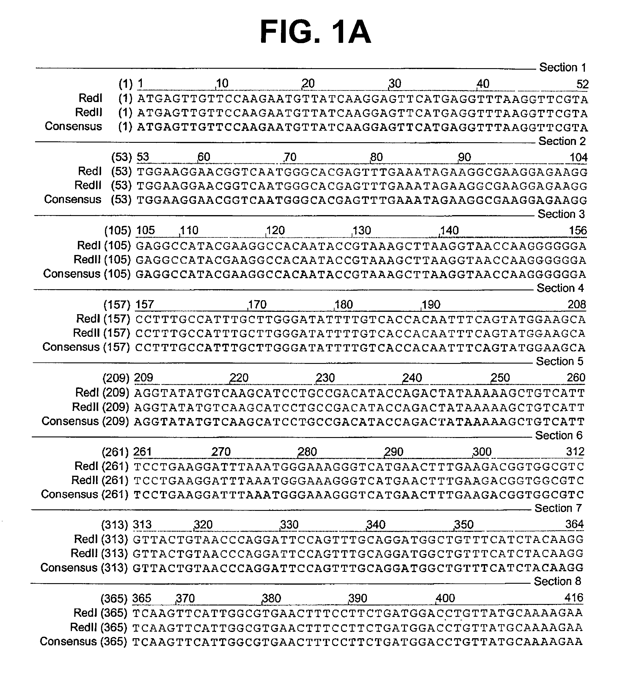
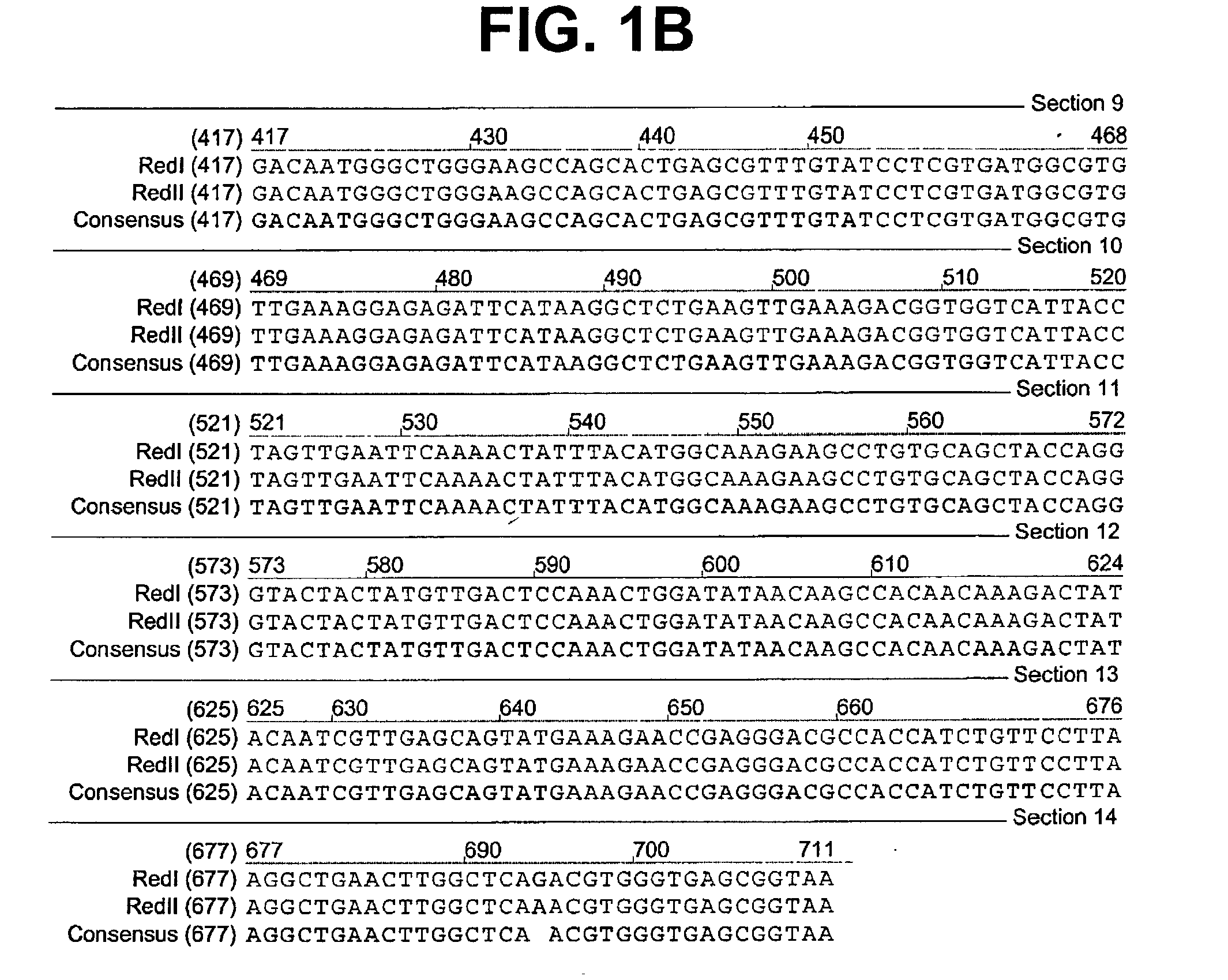
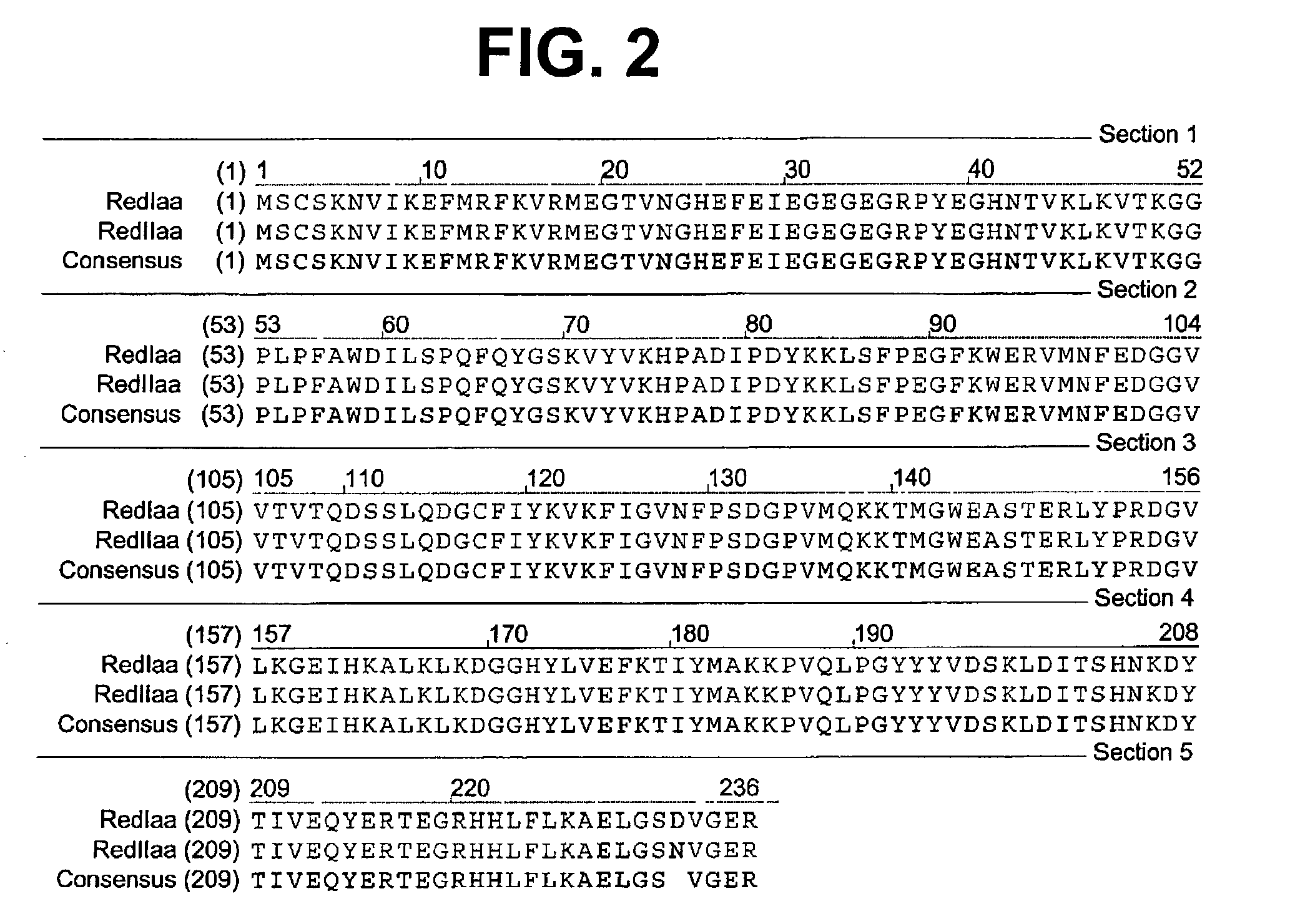
[0076] A colony expressing a wild-type red fluorescent protein (RFP) was isolated from the bacterial colonies generated from the Actinodiscus / Discosoma sp. 1 RNA. This is herein referred to as Ac / DsRFP.

[0077] In addition, a colony expressing a wild-type green fluorescent protein (GFP) was isolated from ...


PUM

PropertyMeasurementUnit
concentrationaaaaaaaaaa
pHaaaaaaaaaa
optical propertyaaaaaaaaaa

Abstract

Provided are four new fluorescent proteins. The proteins were derived from two wild-type fluorescent proteins: a red fluorescent protein (RFP) that was isolated from Actinodiscus or Discosoma sp. 1 and a green fluorescent protein (GFP) isolated from Montastraea cavernosa. Two mutant forms were generated from each wild-type protein. Each of the mutated forms has a higher fluorescence intensity than the respective wild-type form. The mutant forms of the fluorescent proteins allow for more sensitive detection of the fluorescence emitted by the proteins. Additionally, one of the mutant proteins is more resistant to photobleaching than its wild-type protein. The invention also encompasses isolated nucleic acids encoding the mutant forms of the wild-type RFP and GFP.

Description

BIBLIOGRAPHY[0002] Complete bibliographic citations of the references referred to herein by the first author's last name in parentheses can be found in the Bibliography section, immediately preceding the claims.[0003] The invention relates to the field of biochemical assays and reagents. More specifically, this invention relates to fluorescent proteins and to methods for their use.DESCRIPTION OF THE RELATED ART[0004] The number of available fluorescent reporter genes has increased as researchers have isolated genes encoding fluorescent proteins from an increasing variety of organisms and included the genes in cloning cassettes. For example, fluorescent proteins from sea creatures have been used as reporter genes capable of integration into DNA via cloning cassettes. Products of these genes fluoresce under certain wavelengths of light, permitting the tracking of proteins in, e.g., heterologous cells, such as dog and monkey cells. The most commonly used proteins of this nature fluores...

Claims


Application Information

Patent Timeline
Patent Type & Authority Applications(United States)
IPC IPC(8): A01K67/027C07K14/435C12N15/85
CPCA01K67/0275C12N15/8509C07K14/43595A01K2227/40C07K14/47C12N5/16C12N13/00
Inventor GIBBS, PATRICK D.L.CARTER, ROBERT W.SCHMALE, MICHAEL C.
Owner MIAMI UNIVERISTY OF
Why Eureka
Try Eureka