Microvascular free flaps for local or systemic delivery

a microvascular and free flap technology, applied in the field of vertebrate tissue explantation, can solve the problems of limited availability of most donor organs, and achieve the effect of avoiding the immunologic complexity of xenotransplantation and minimal or no finctional loss

Inactive Publication Date: 2004-12-09
NEW YORK UNIV
View PDF14 Cites 7 Cited by
  • Summary
  • Abstract
  • Description
  • Claims
  • Application Information

AI Technical Summary

Benefits of technology

0027] The compositions and methods of the invention are particularly advantageous in that they avoid many of the problems associated with viral transfection of tissue in vivo. Using the methods of the invention, explanted microvascular flaps (or beds) can be modified ex vivo, via genetic transfection or introduction of cells, then reattached to the native circulation. In the case of genetically modified tissues, this enables high-level localized expression of the nucleic acid encoding a product of interest in cells of the explanted flap (or bed), with little or no collateral transfection occurring in other tissues. Current methods of human gene therapy, on the other hand, require systemic administration of a nucleic acid of interest (in vivo) or removal of isolated cells for modification (ex vivo) and subsequent re-infusion. Furthermore, the methods of the invention are reversible, in that the flap can be removed, in the event that the recipient displays an adv

Problems solved by technology

Furthermore, most donor organ

Method used


Image

  • Microvascular free flaps for local or systemic delivery
  • Microvascular free flaps for local or systemic delivery
  • Microvascular free flaps for local or systemic delivery

Examples


example 1

6. EXAMPLE 1

Ex Vivo Transfection of Microvascular Free Flaps

[0191] 6.1. Introduction

[0192] Gene therapy using viral vectors holds great clinical promise but has been limited by difficulties in developing targeted, high-level gene expression with acceptable host toxicity.

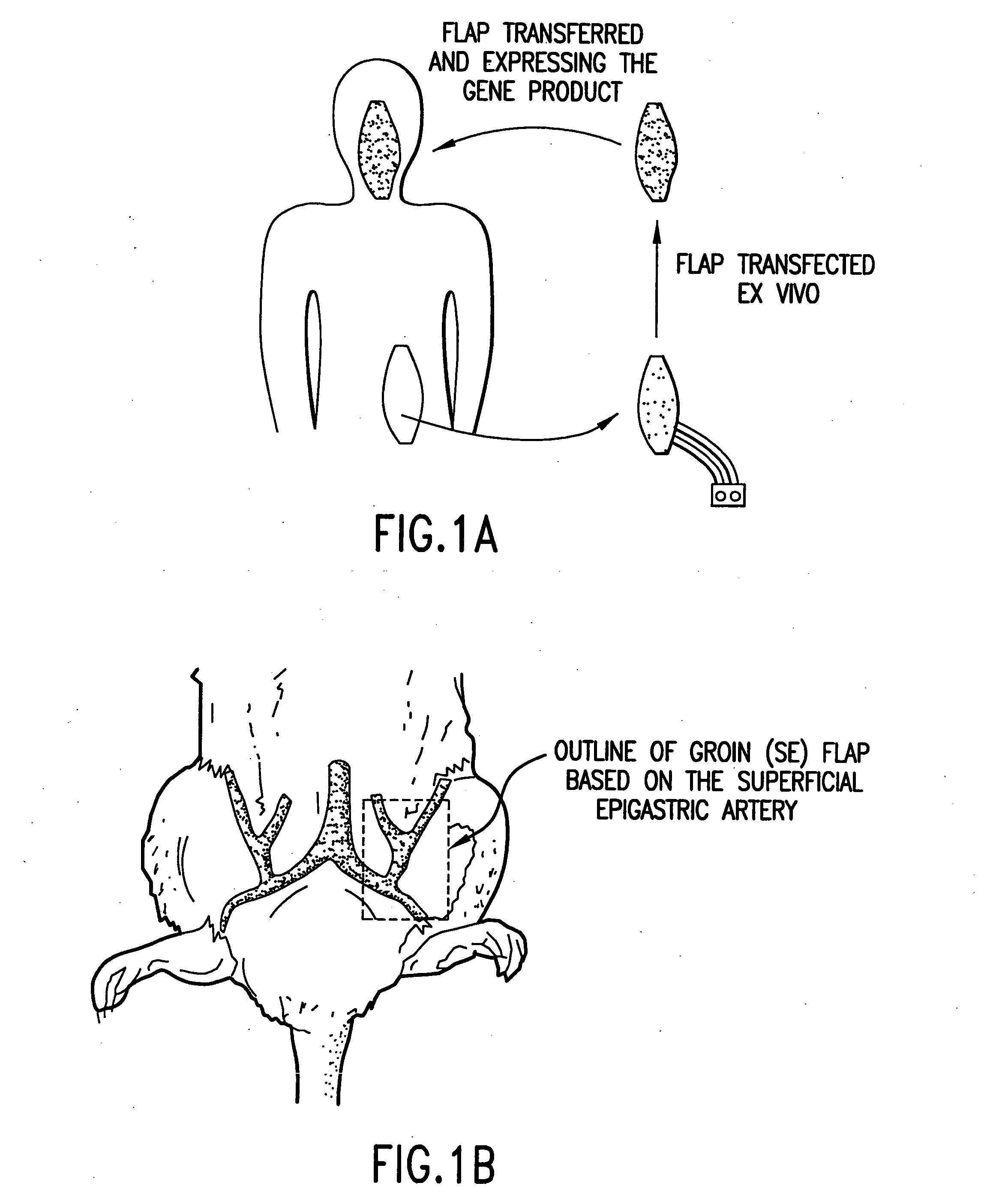
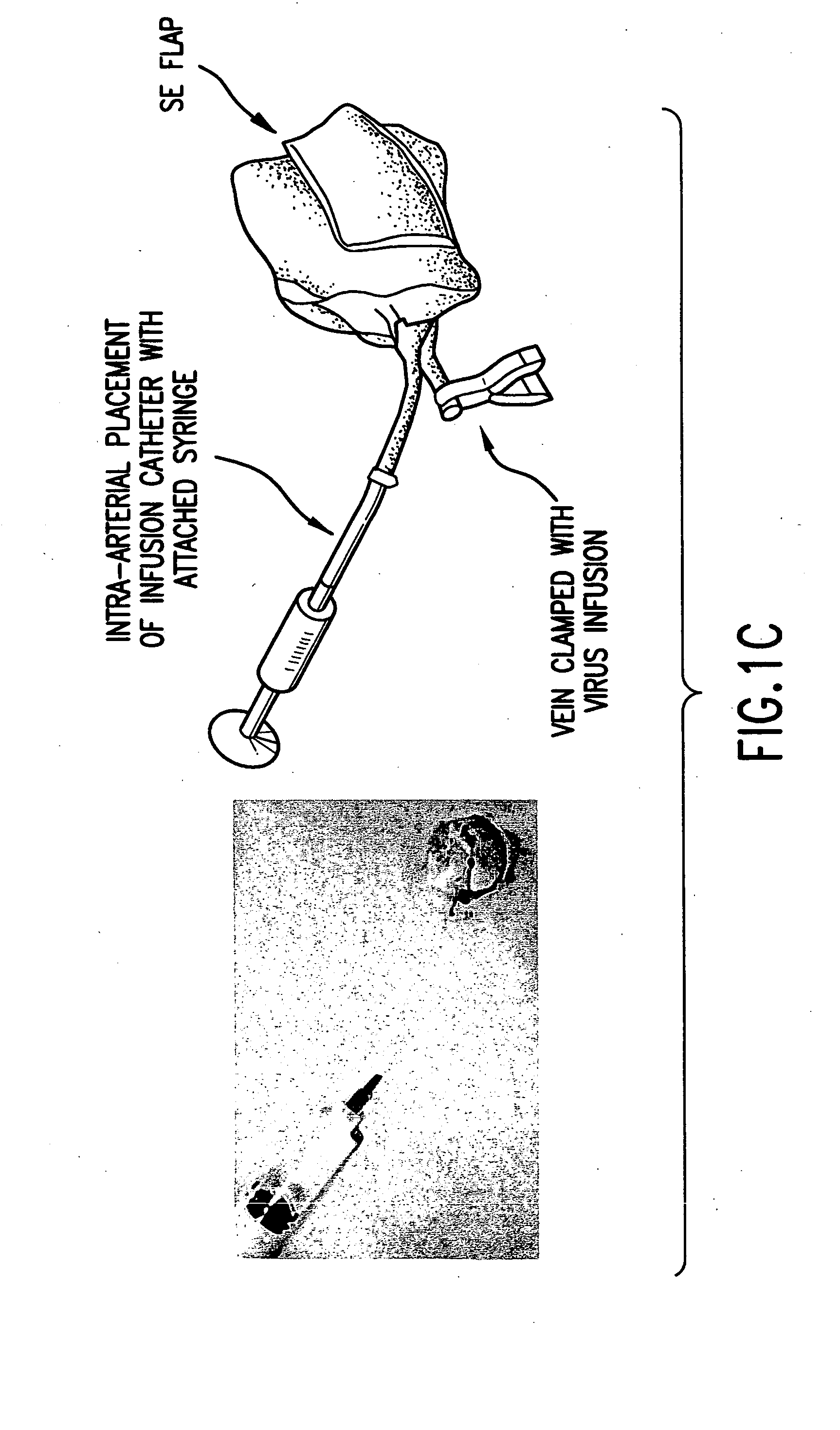
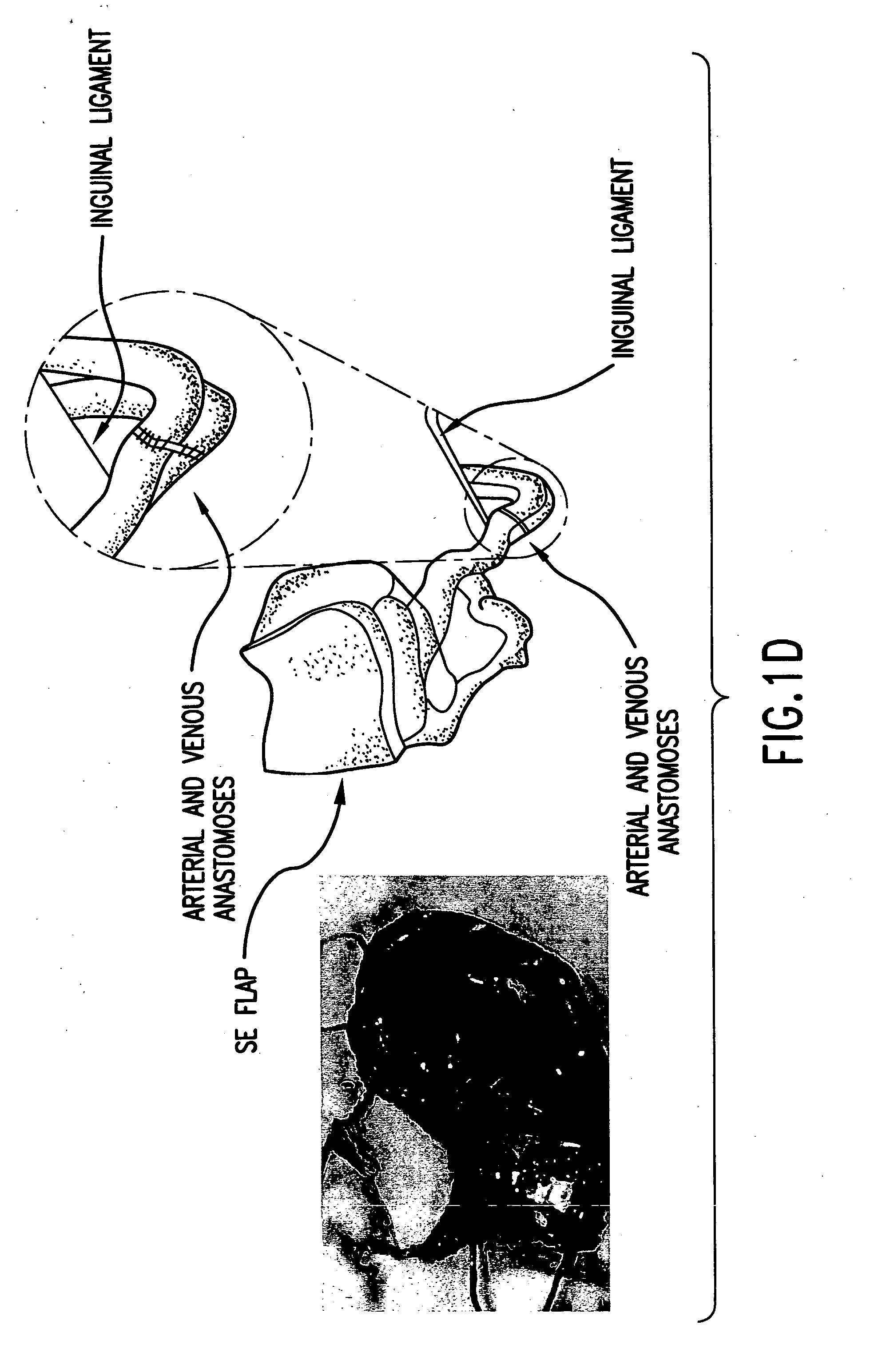
[0193] The following example demonstrates a method of delivery of a nucleic acid encoding a product of interest that avoids many of the problems associated with viral transfection. Using a rat model, explanted microvascular free flaps were transfected ex vivo, flushed, and reattached to the native circulation using microvascular techniques. A nucleic acid encoding .beta.-galactosidase (.beta.-gal) as a reporter gene was used to demonstrate delivery of a product of interest according to the methods of the invention. Transfection was performed using an adenoviral vector containing the .beta.-galactosidase (.beta.-gal) reporter gene driven by the CMV promoter.

[0194] High regional expression of the .beta.-gal gene was se...

example 2

7. EXAMPLE 2

Transfection of Microvascular Free Flaps with a Nucleic Acid to Ameliorate the Effects Diabetes Mellitus

[0216] 7.1. Introduction

[0217] In this example, a rat superficial epigastric (SE) flap is genetically modified ex vivo with a nucleic acid encoding a therapeutic molecule of interest, i.e., the nucleic acid encoding proinsulin. The flap is re-implanted into the donor, where it functions as a neo-organ that delivers insulin following re-anastomosis.

[0218] The rat superficial epigastric flap is used as a model, principally because of its reproducibility and technical ease (Perry et al., 1984, Plast. and Recon. Surg. 74(3): 410-3). As will be understood by those skilled in the art, animal models (e.g., the mouse flap model (Cooley et al., 1998, Microsurgery 18(5): 320-3,), can be used to demonstrate the advantageous utility of the microvascular free flap method of the invention to deliver insulin systemically to an animal in need thereof The mouse model is advantageous be...

example 3

8. EXAMPLE 3

Transfection of Epigastric Free Flaps with IL-12

[0231] 8.1. Introduction

[0232] In this example, a rat tumor model is used to confirm that a genetically modified pigastric free flap can act as a delivery vehicle for localized gene therapy with emonstrable antitumor effect. A rat subcutaneous tumor model is employed and the rat istiocytoma cell line AK-5 is used as the tumor source (Nandakumar et al., 1997, Cytokines Cell. Mol. Ther. 3(4): 225-32). Prior studies have demonstrated that this tumor is exquisitely sensitive to local IL-12 therapy when delivered via injection. The methods exemplified herein may also be applied to a mouse flap model (Cooley et al., 1998, Microsurgery 18(5): 320-3). As will be understood by those skilled in the art, animal models (e.g., the mouse flap model), can be used to demonstrate the advantageous utility of the microvascular free flap of the invention to act as a delivery vehicle for localized gene therapy. The mouse model is advantageous b...


PUM

PropertyMeasurementUnit
Timeaaaaaaaaaa
Timeaaaaaaaaaa
Timeaaaaaaaaaa

Abstract

The present invention relates to methods of ex-vivo modification of mammalian tissue, via genetic transformation or introduction of cells, followed by implantation of the modified tissue into a patient in need thereof. Preferably, the tissue is microvascular free flap (or microvascular bed) tissue. A tissue explant is detached from the native circulation of a donor, transfected ex vivo, and then attached (anastomosed) to a recipient, either the donor or another patient. In a preferred embodiment, the mammalian tissue is human tissue and the patient is a human patient. Transfection with a nucleic acid encoding a product of interest is performed by contacting the selected tissue with a vector, preferably a viral vector, most preferably an adenoviral vector, that comprises the nucleic acid encoding the product of interest. The nucleic acid encoding the product of interest is driven by regulatory element such as an inducible, constitutive or cell-specific promoter, preferably an inducible or constitutive promoter. After genetic transformation of the selected tissue, the tissue is flushed to remove the vector not incorporated into the cells of the tissue. The tissue is then attached to the native circulation of the recipient using microvascular techniques. In one aspect, the invention provides methods of local delivery of a product (protein) of interest. In another aspect, the invention provides methods of systemic delivery of a product of interest. In yet another aspect, the invention provides methods of both local and systemic delivery of a product of interest. In yet another aspect, the invention provides methods for producing a "neo-organ," i.e., a non-naturally occurring vascularized tissue that provides a function of a gland or organ, or that supplements the function of a gland or organ, and that delivers locally or systemically a product of interest to a patient in need thereof.

Description

[0001] This application claims priority benefits of application No. 60 / 289,452 filed May 7, 2001, the entire disclosure of which is incorporated herein by reference in its entirety.[0002] 1. TECHNICAL FIELD[0003] The present invention relates to methods of ex-vivo modification of vertebrate tissue, via genetic transformation or introduction of cells, for local or systemic delivery of a therapeutic product of interest. The present invention also relates to methods for producing a "neo-organ," i.e., a non-naturally occurring vascularized tissue that provides a function of a gland or organ, or that supplements the function of a gland or organ.2. BACKGROUND OF THE INVENTION[0004] Gene therapy using viral vectors holds great clinical promise but has been limited by difficulties in developing targeted, high-level gene expression with acceptable host toxicity. Human gene therapy has been a disappointment in clinical trials and in some gene therapy trials serious concerns regarding safety h...

Claims


Application Information

Patent Timeline
IPC IPC(8): A61K35/12A61K35/44A61K38/28A61K48/00
CPCA61K38/28A61K48/00A61K48/0075A61K38/208A61K38/39A61K35/44A61K2300/00
Inventor GURTNER, GEOFFREY C.
Owner NEW YORK UNIV
Why Eureka
Try Eureka