
Biological sample component purification and differential display

a biological sample and component technology, applied in the field of biological sample component purification and differential display, can solve problems such as affecting their ability, and achieve the effect of reducing the complexity of biological samples

Inactive Publication Date: 2005-01-06
CHIRON CORP
View PDF21 Cites 13 Cited by
  • Summary
  • Abstract
  • Description
  • Claims
  • Application Information

AI Technical Summary

Benefits of technology

Affinity supports in accordance with the present invention may be used in a variety of techniques and apparatuses to achieve improved separations of complex biological samples and thereby enhance the results of biological sample component resolutions, fractionations, enrichments, purifications, expression product determinations and comparisons, and other biological sample processing and analysis techniques. In addition, the affinity supports may be included in kits useful in processing biological samples for applications such as novel protein purification.
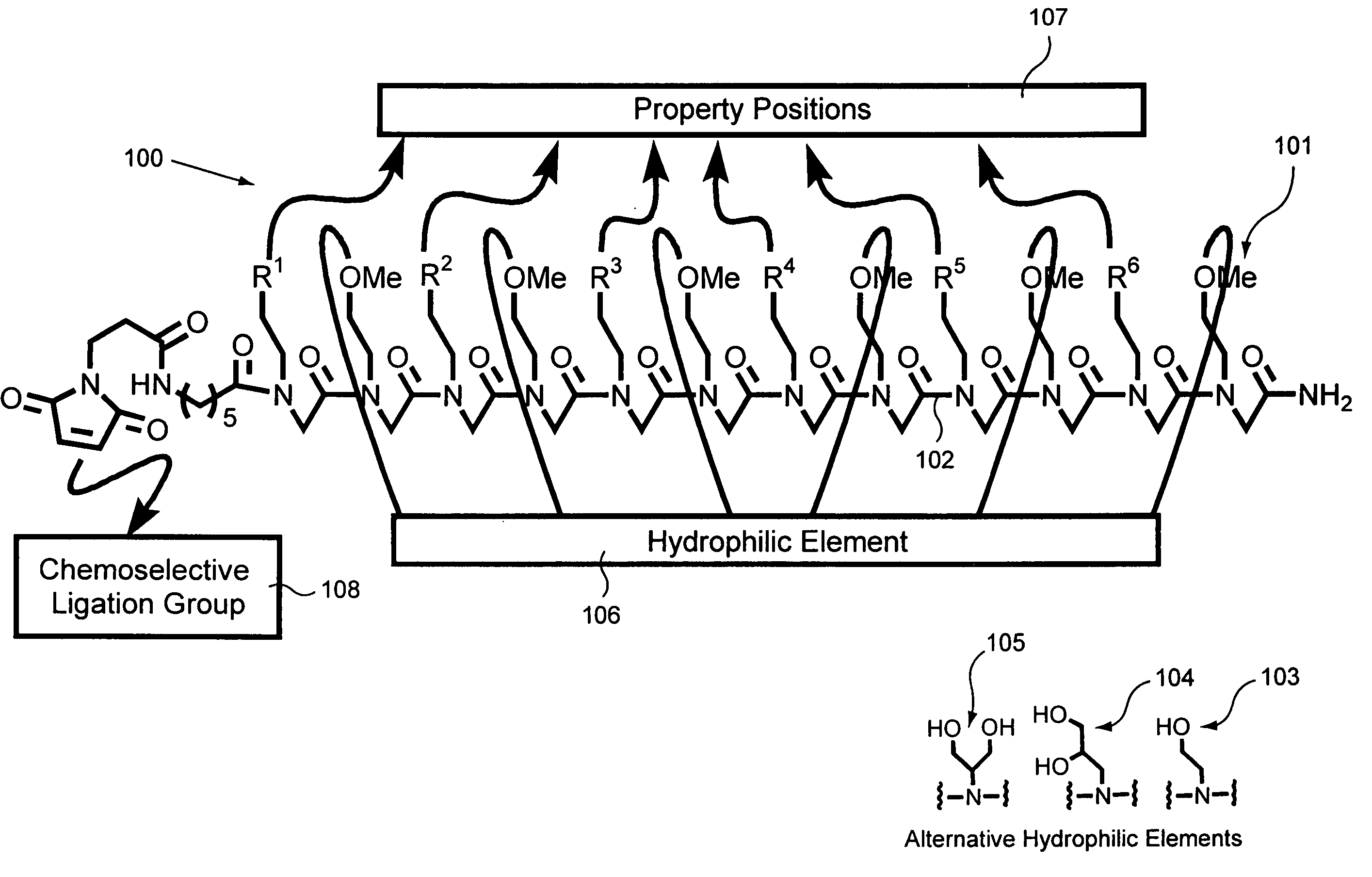
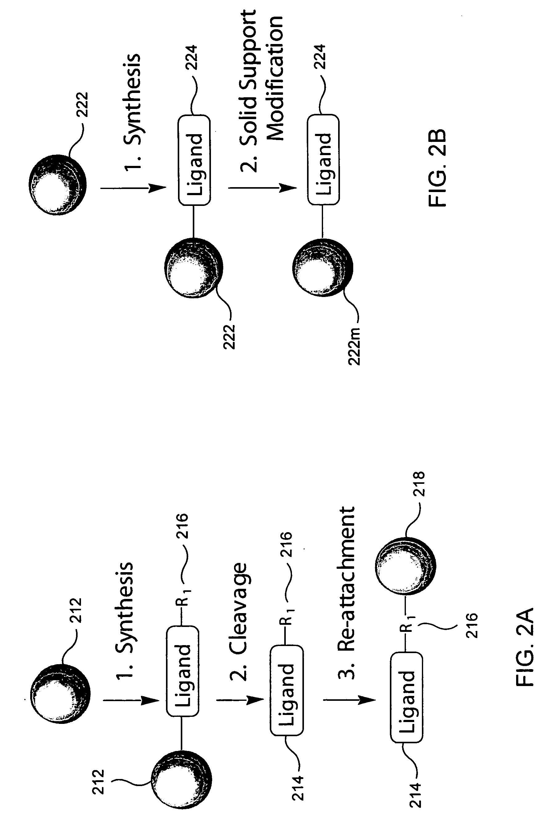
In other embodiments, the invention provides, methods of comparing biological sample phenotypes, reducing complexity of biological samples, massively parallel processing a biological sample on a number of affinity supports both one- and two-dimensionally, and materials and kits incorporating affinity supports useful in accomplishing these methods. Also provided are methods of making affinity supports in accordance with the present invention, in particular supports including peptoids having both affinity property groups and hydrophilic groups pendent from their peptoid backbones and coupled to hydrophilic matrixes.

Problems solved by technology

Such structures may aggregate to form micelles thus detracting from their ability to achieve separations and / or couple efficiently with the substrate.

Method used


Image

  • Biological sample component purification and differential display
  • Biological sample component purification and differential display
  • Biological sample component purification and differential display

Examples


example 1

Preparation of a Peptoid on a Hydrophilic Support

Peptoids were prepared on Rink amide polystyrene resin as illustrated and described in FIG. 6, part A, Peptoid Synthesis. The synthesis procedure is also reported in Figliozzi, G. M., Goldsmith, R., Ng, S. C., Banville, S. C., Zuckermann, R. N. Methods Enzymol. 1996, 267, 437-447, the disclosure of which is incorporated by reference herein for all purposes.

Before cleavage, the peptoids on Rink resin were treated with N-FMOC-aminohexanoic acid (0.4M), HOBT (0.4M), DIC (0.44M) at 35° C. for 2 hr. Double coupling was performed when the ninhydrin test exhibit blue colored beads. After piperidine treatment to remove the FMOC protecting group, the beads were treated with 3-maleimido propionic acid (0.4M), HOBT (0.4M), DIC (0.44M) for 1 hr. at 35° C. After cleavage from the resin using 95% TFA [v / v] in H2O the resulting solution was filtered, mixed with H2O, frozen and lyophilized. The resulting product was dissolved in 100% AcOH, frozen...

example 2

Evaluation of Fabricated Hydrophilic Affinity Supports

Each solid phase attached peptoid was packed in a column and loaded with a cell lysate. After a certain period, the column was washed with the loading buffer. The resin bound proteins were eluted using a variety of elution buffers. The fractions collected were analyzed using polyacrylamide gel electrophoresis.

The versatility of these materials was evaluated using four criteria: reproducibility, binding capacity, enrichment and specificity. The structures of the various peptoids used in these evaluation are found in FIGS. 3A-C.

FIG. 7, shows the elution product reproducibility of peptoid / sepharose column after three loadings, washings and elutions of the HeLa cell lysate. The gel shows a high degree of reproducibility.

FIG. 8 shows the binding capacity of peptoid sepharose columns EB8224 and EB8225 toward protein sets of a HeLa cell lysate. EB8224 bound 7% of the cell lysate, while EB8225 bound 11% of the cell lysate. This dem...

example 3

Identification of a Support Ligand that Selectively Binds

Four peptoid-based affinity supports prepared in accordance with the present invention were used to purify a protein from a crude cell lysate. The four columns used in this example, identified as 01, 02, 03 and 04 in FIG. 10, were the columns identified as EB8201, EB8202, EB8203 and EB8204, respectively, in FIGS. 3A and 3C. A peptoid that binds selectively to the protein FGF (Fibroblast Growth Factor) was located. Each different column was loaded with a FGF-containing cell lysate and run using the cationic conditions described above in Table 1. The flow through fraction (FT) from the column and the initially bound and then eluted fraction (Eluate) were subjected to gel electrophoresis together with a FGF standard. The peptoid ligand (FIG. 4b, lane 6; bFGF) that bound selectively to FGF (FIG. 4b, lane 10) was determined, as shown in FIG. 10.


PUM

PropertyMeasurementUnit
affinityaaaaaaaaaa
hydrophilicaaaaaaaaaa
binding affinityaaaaaaaaaa

Abstract

Provided are affinity support materials having intermediate binding affinity for biological samples. Among the materials provided by the present invention are hydrophilic solid supports composed of hydrophilic ligands coupled to hydrophilic matrixes which are compatible with biological samples, for example, a cell line, a biological fluid such as blood, or a tissue cell lysate. The ligands may include affinity property groups and hydrophilic groups pendent from a backbone, and be configured to at least partially resolve components of a biological sample. Affinity supports in accordance with the present invention may be used in a variety of techniques and apparatuses to achieve improved separations of complex biological samples and thereby enhance the results of biological sample component fractionations, enrichments, purifications, expression product determinations and comparisons, and other biological sample processing techniques. In addition, the affinity supports may be included in kits useful in processing biological samples.

Description

BACKGROUND OF THE INVENTION The present invention relates to techniques and materials for the purification of biological samples and the differential display and analysis of components of such biological samples. In one embodiment, the invention is directed to protein purification and proteome differential display. Current methodologies for purifying biological sample components (e.g., protein, nucleic acid, etc.) rely on a few commercially available hydrophilic solid supports. Conventional chromatography supports include anionic exchange supports, such as diethylaminoethyl (DEAE) Sepharose, etc., cationic exchange supports, for example having carboxymethyl functional groups, reverse phase and hydrophobic interaction supports. These materials are able to distinguish between biological sample components, such as proteins, only very generally and are therefore limited in their ability to separate biological sample components, particularly proteins of similar anionic, cationic, etc. ...

Claims


Application Information

Patent Timeline
Patent Type & Authority Applications(United States)
IPC IPC(8): B01D15/38B01J20/286B01J20/32G01N33/53G01N33/543
CPCB01D15/3804B01J20/286Y10T436/25375Y10S435/814B01J20/3242
Inventor ZUCKERMANN, RONALD N.BEAUSOLEIL, ERICWACHOWICZ, MATTHEWKOTHAKOTA, SRINIVAS
Owner CHIRON CORP
Learn More