

Non-invasive measurement of blood glucose using retinal imaging
a retinal imaging and blood glucose technology, applied in the field of blood glucose in vivo measurement, can solve the problems of high cost of glucose testing supplies, blood drawn for analysis, and many patients not testing their blood as often as recommended, so as to achieve accurate blood glucose determination
- Summary
- Abstract
- Description
- Claims
- Application Information
AI Technical Summary
Benefits of technology
Problems solved by technology
Method used
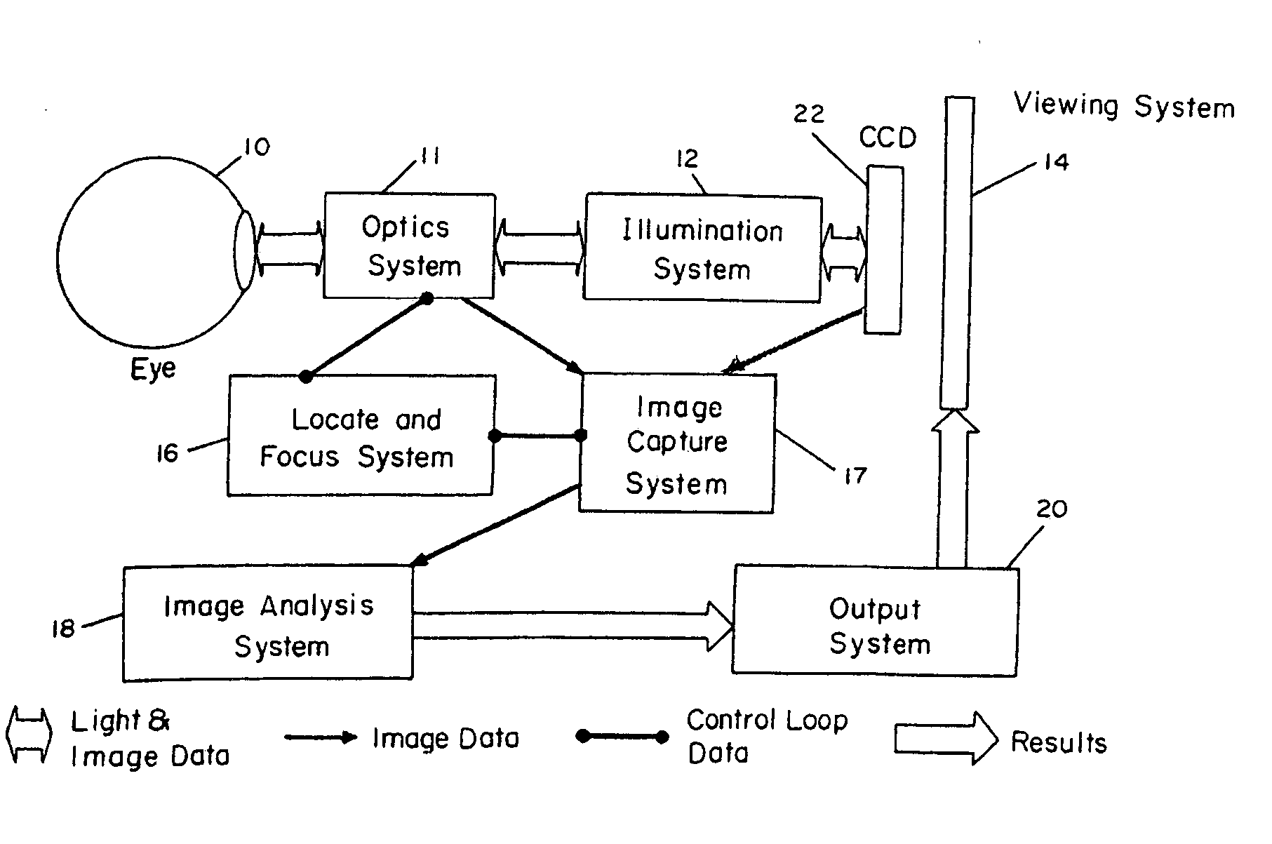
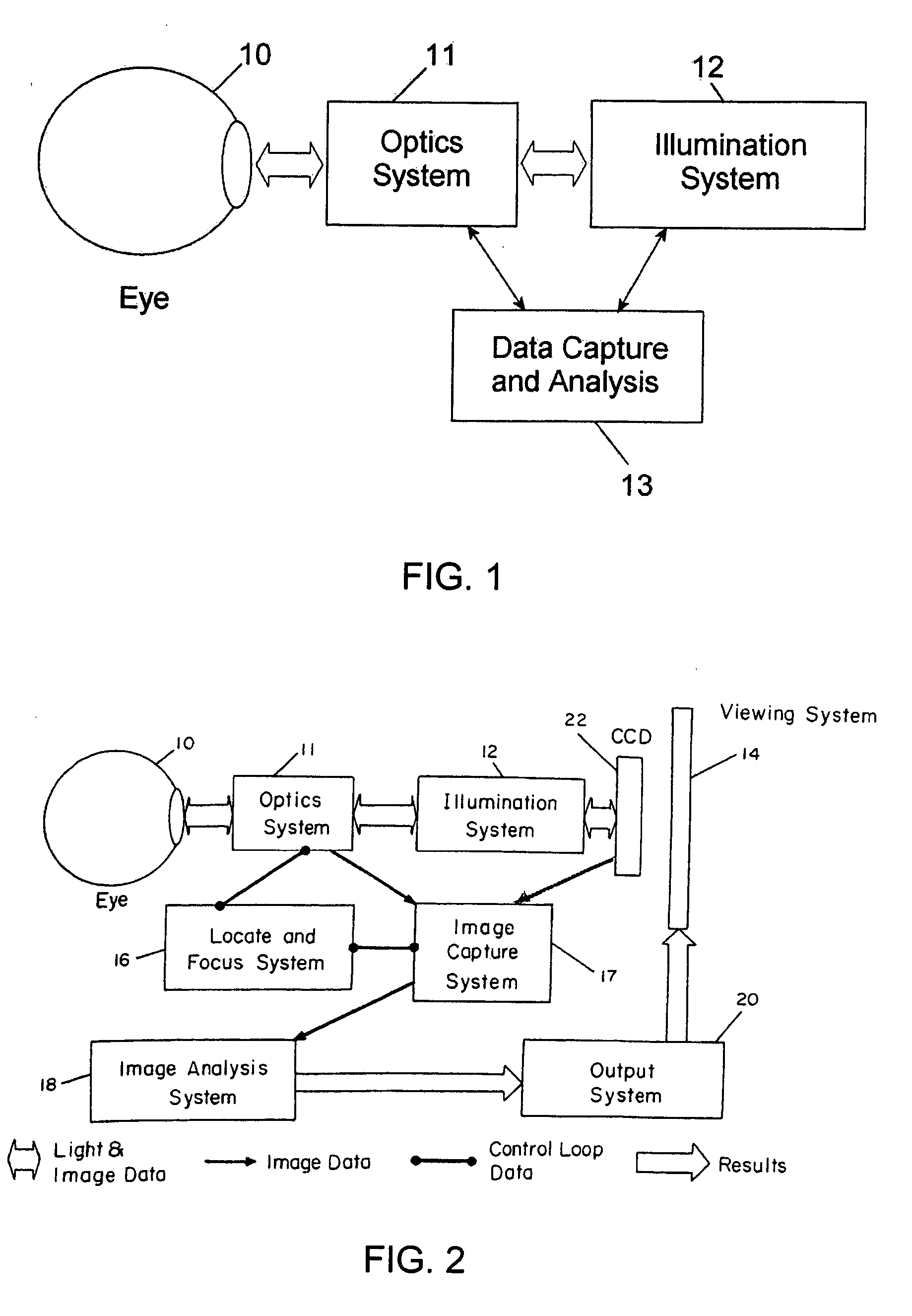
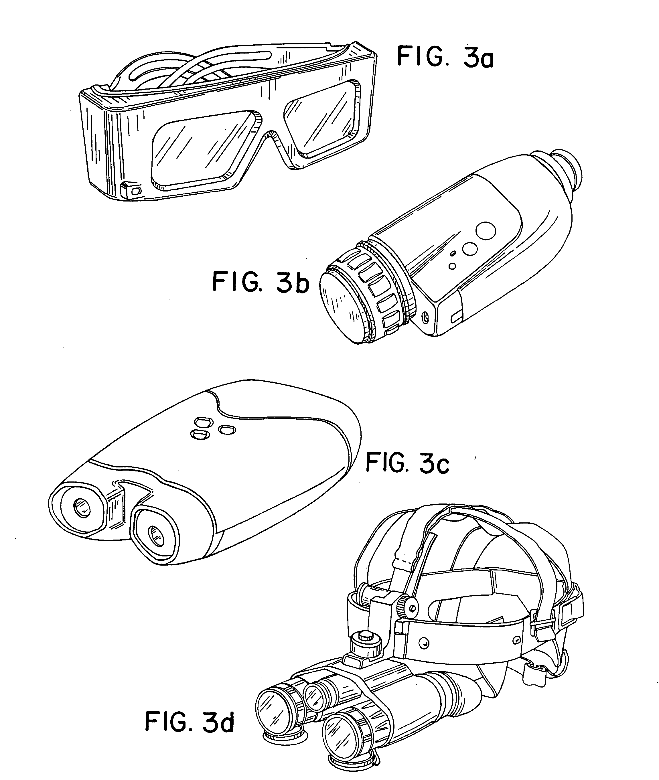
Image

Examples
Embodiment Construction
[0035] Rhodopsin is the visual pigment contained in the rods (that allow for dim vision) and cone visual pigment is contained in the cones of the retina (that allow for central and color vision). The outer segments of the rods and cones contain large amounts of visual pigment, stacked in layers lying perpendicular to the light incoming through the pupil. As visual pigment absorbs light, it breaks down (bleaches) into intermediate molecular forms and initiates a signal that proceeds down a tract of nerve tissue to the brain, allowing for the sensation of sight. During normal vision this bleaching process occurs continuously. Light that reacts with the visual pigments causes a breakdown of those pigments. This phenomenon is termed bleaching, since the retinal tissue loses its color content when a light is directed onto it. In addition, regeneration of the visual pigments occurs at all times, even during the bleaching process. Rod visual pigment absorbs light energy in a broad band cen...
PUM
 Abstract
Description
Claims
Application Information