

Cardiac muscle regeneration using mesenchymal stem cells
a technology of mesenchymal stem cells and cardiac muscle, applied in the direction of genetically modified cells, skeletal/connective tissue cells, drug compositions, etc., can solve the problems of high mortality due to this disorder, improper heart valve function, and acellular valves, etc., to shorten the time required for complete differentiation
- Summary
- Abstract
- Description
- Claims
- Application Information
AI Technical Summary
Benefits of technology
Problems solved by technology
Method used
Image

Examples
example 1
[0040] Implantation of MSCs in Normal Cardiac Muscle
[0041] In using MSCs, it is desirable to maintain cell-cell contact in vivo for the conversion of MSCs to the muscle lineage. Environmental signals identified above act in concert with mechanical and electrical signaling in vivo to lead to cardiac differentiation.
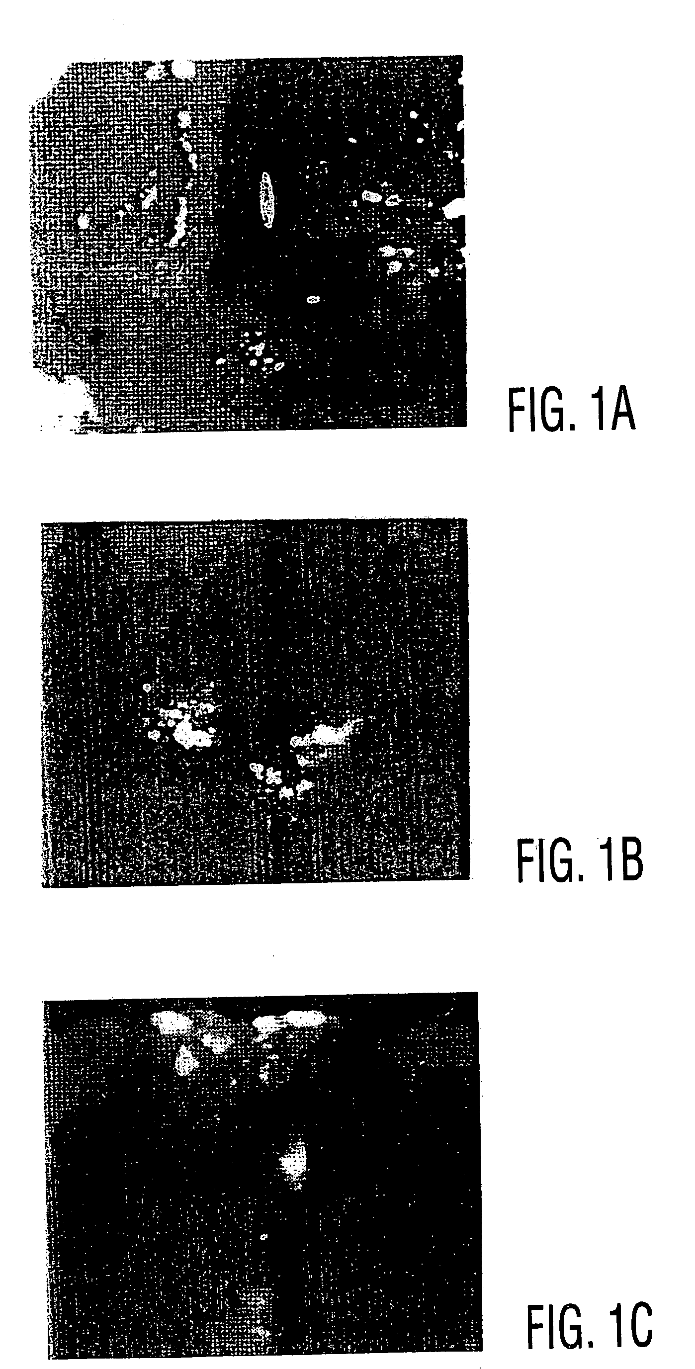
[0042] Primary human MSCs (hMSCs) are introduced into athymic rat myocardial tissue by direct injection. The integration of implanted cells, their subsequent differentiation, formation of junctions with cardiac cells, and their long-term survival are characterized with light microscopy, histology, confocal immunofluorescence microscopy, electron microscopy and in situ hybridization.
[0043] Whether human MSCs are appropriately grafted into cardiac muscle of athymic rats (strain HSD:RH-RNU / RNU), which lack the immune responses necessary to destroy many foreign cells, is also examined.
[0044] Rat MSCs are grafted into the heart muscles of rats. To analyze the injected cells...
example 2
[0052] Regeneration of Heart Valves Using MSCs
[0053] Xenograft or homograft valves are made acellular by freeze-drying, which leads to cellular death, or by enzymatic treatment followed by detergent extraction of cells and cell debris. This latter approach was taken by Vesely and coworkers with porcine valves to be repopulated with dermal or aortic fibroblasts. Curtil, et al. 1997 used a freeze-dried porcine valve and attempted repopulation of the valve with human fibroblasts and endothelial cells. These studies were preliminary and limited to short term studies in vitro.
[0054] The acellular valve to be populated by autologous hMSCs is incubated with culture expanded hMSCs in a tumbling vessel to ensure loading of cells to all valve surfaces. The valve is then cultured with the hMSCs for 1-2 weeks to allow the hMSCs to infiltrate and repopulate the valve. Within the culture vessel, the valve is then attached to a pump to allow the actuation of the valve leaflets and simulate the p...
example 3
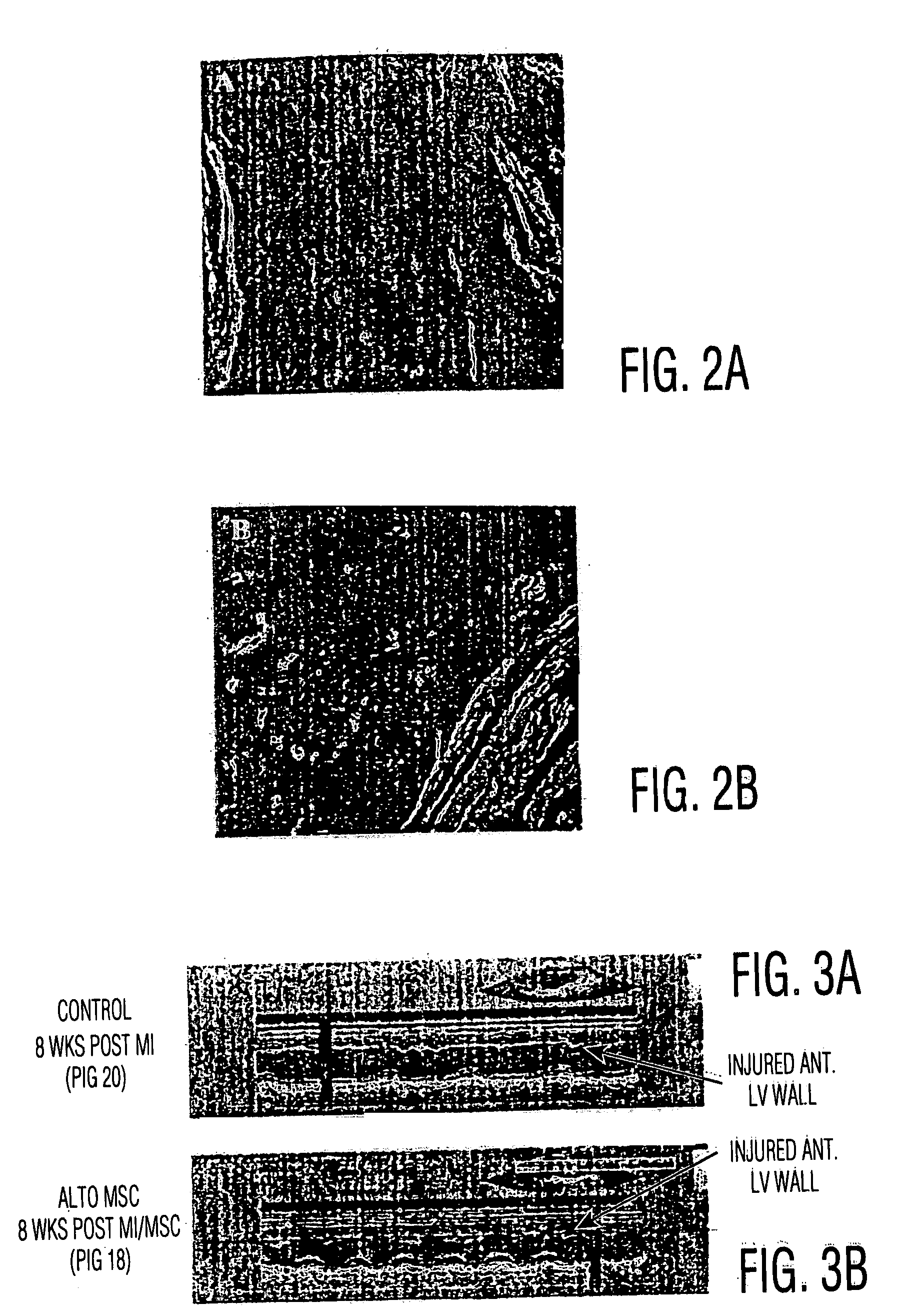
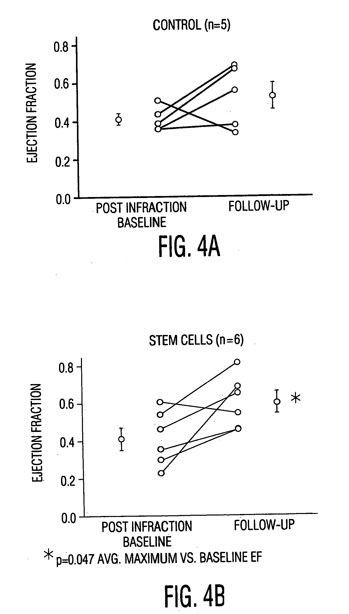
[0056] MSC Engraftment in Rat MI Model: Direct Injection Vs. Systemic Delivery
[0057] Myocardial infarction was produced in Fisher rats as follows:
[0058] Fisher rats were given a cocktail of Ketamine / Xylazine / Acepromazine (8.5 mg / 1.7 mg / 0.3 mg I.P.) The depth of anesthesia was assessed using a toe-pinch and eye-blink reflexes. When a surgical plane of anesthesia was achieved, endotracheal intubation was performed and the animal placed on 1.0% Isoflorane. Positive-pressure breathing was provided throughout the procedure by means of the Engler ADS 1000 small animal ventilator. A left thoracotomy was performed and the pericardium opened. A 6-0 silk ligature snare was then placed around the left anterior descending (LAD) coronary artery at a location distal to the first diagonal branch. A brief (30 sec) LAD test occlusion is performed to insure that a modest region of ischemia is procued, involving a limited region of the anterior free wall and septum. Ischemia is confirmed by characte...
PUM
| Property | Measurement | Unit |
|---|---|---|
| Volume | aaaaa | aaaaa |
| Volume | aaaaa | aaaaa |
| Volume | aaaaa | aaaaa |
Abstract
Description
Claims
Application Information