Method and device for manufacturing tissue section

a tissue and manufacturing technology, applied in the field of tissue manufacturing, can solve the problems of difficult individual recognition, limited method, and risk of infection of frozen organism specimens, and achieve the effects of uniform thickness, easy individual recognition, and stab manufacturing

Active Publication Date: 2005-06-16
SAKURA SEIKI
View PDF5 Cites 11 Cited by
  • Summary
  • Abstract
  • Description
  • Claims
  • Application Information

AI Technical Summary

Benefits of technology

[0034] As described above, since the entire tissue section can be rapidly adhered on the film running apart from the organism specimen, the film is not pressed onto the organism specimen, so that no tissue cells of the organism specimen are broken and the tissue section having uniform thickness can be stably manufactured.
[0035] Further, since the adhesion of the tissue section is controlled on the basis of the temperature difference between the film and the organism specimen, no adhesive is applied to the one side of the film so that no tissue cells are badly influenced and transformed by adhesive. In the present invention, the tissue section is immediately adhered onto the film after the tissue section is cut off from the organism specimen; even if a plurality of the tissue sections are sliced, the tissue sections can be adhered onto the film in slicing order, so that the tissue section can be easily individually recognized.

Problems solved by technology

Further, there is a risk of infection from the frozen organism specimen 12.
Further, if air is left in a space between the transparent film and the organism specimen, creases are easily formed in the tissue section cut off from the organism specimen, so that it is necessary to discharge the air by pressing the transparent film onto the organism specimen with a prescribed force.
In the method such that the tissue section is scooped from water by the transparent film, frozen organism specimens cannot be used, so the method is limited to organism specimens embedded by a hydrophobic embedding agent, e.g., paraffin.
Further, in the case of using the embedded organism specimens embedded by the hydrophobic embedding agent, e.g., paraffin, if a plurality of tissue sections are dipped into water, it is difficult to individually recognize them, so one tissue section is dipped into the water after the prior tissue section is scooped by the transparent film.

Method used


Image

  • Method and device for manufacturing tissue section
  • Method and device for manufacturing tissue section
  • Method and device for manufacturing tissue section

Examples


experiment 1

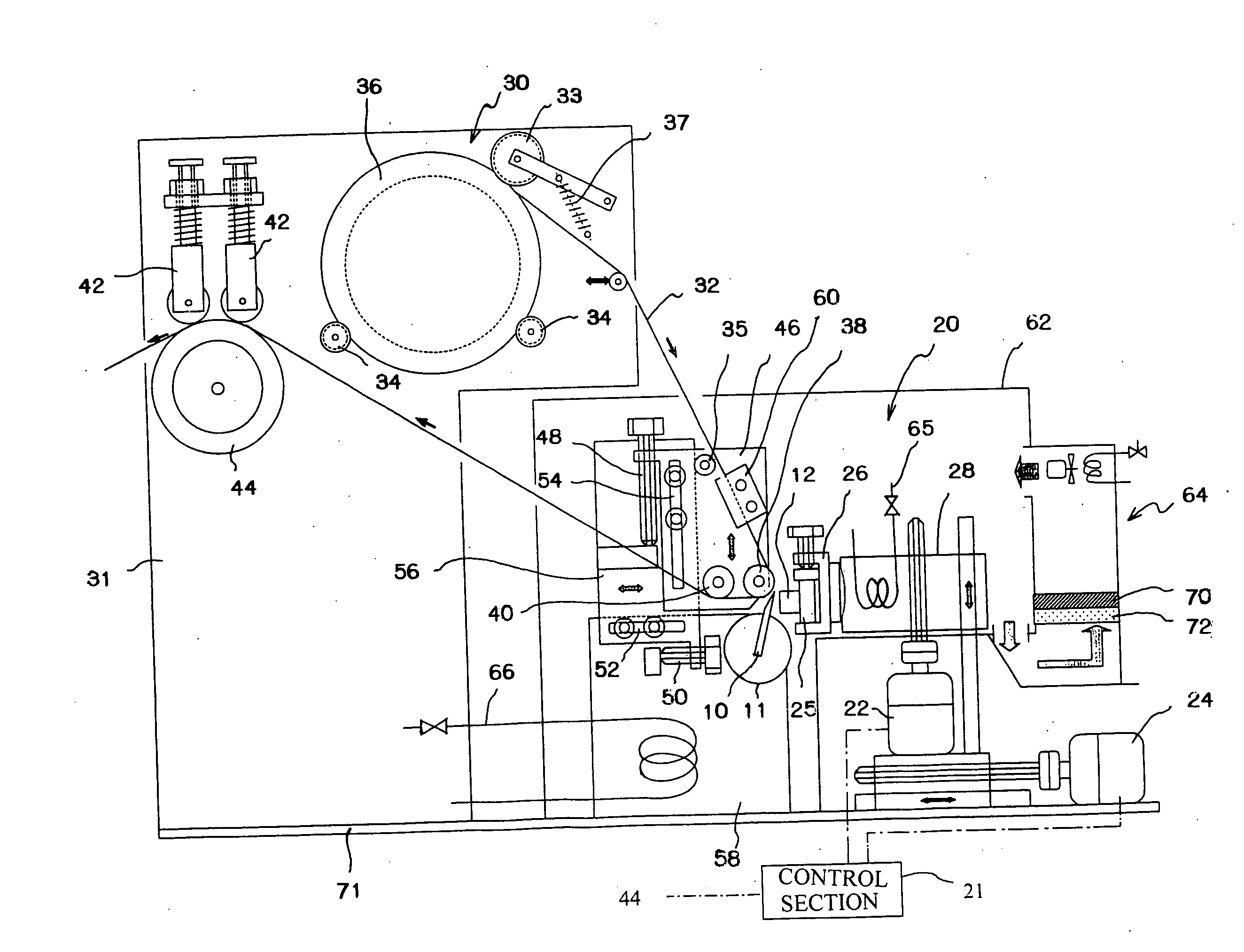
[0105] In the device shown in FIG. 1, the tissue sections 16 were made by slicing the frozen organism specimen 12, which was cut off from a pig lung, liver or muscle.

[0106] The tape 32 used in the device shown in FIG. 1 was the transparent film tape 32, which was made of cellulose acetate and whose width was 24 mm; the organism specimen 12 was made by the steps of: cutting a piece off from a pig lung, liver or muscle; embedding the cut piece in carboxymethylcellulose as an ice crystal-inhibitor, and freezing the embedded piece.

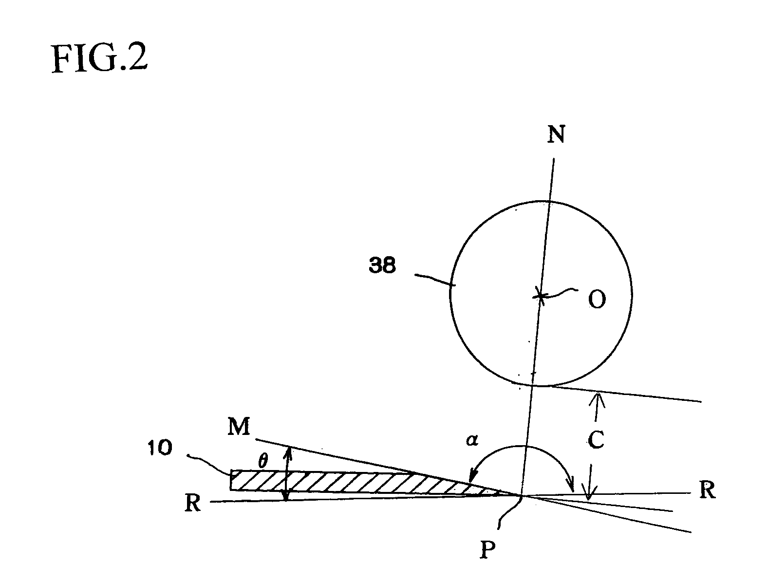
[0107] The frozen organism specimen 12 was fixed to the jig 25, on which carboxymethylcellulose was applied, then the jig 25, to which the frozen organism specimen 12 had been fixed, was fixed to the clamper 26 of the holding section 28. The cylinder 11, to which the knife 10 was attached, was turned until the angle θ (see FIG. 2) between the slicing surface of the frozen organism specimen 12 fixed to the holding section 28 and the cutting surface of the kni...

experiment 2

[0113] In this experiment, the speed of slicing the tissue sections 16, whose thickness was 5 μm, was 60 times / minute, but other conditions were the same as those of the experiment 1; the ratio (Vt / Vs) of the running speed (Vt) of the tape 32 to the moving speed (Vs) of the holding section 28 was 0.9 as well as the experiment 1. Front ends of some tissue sections 16 were broken, but they were usable as microscopic samples.

experiment 3

[0114] In this experiment, the distance C shown in FIG. 2 between the close roller 38 and the frozen organism specimen 12 (see FIG. 2) to 2 mm, but other conditions were the same as those of the experiment 1. Marks existed in some tissue sections 16, but they were accepted.


PUM

PropertyMeasurementUnit
temperatureaaaaaaaaaa
melting pointaaaaaaaaaa

Abstract

The method for manufacturing a tissue section, which is used for a sample for observation by a microscope, comprises the steps of slicing an organism specimen (12), whose form has been fixed by freezing or by using an embedding agent, along a slicing surface, characterized by: adjusting a distance between the slicing surface of the organism specimen (12) and one side of a tape (32) and a temperature difference between the organism specimen (12) and the tape (32) when the slicing of the organism specimen (12) is started, thereby a tip part of the tissue section (16) curling to the outside of the slicing surface of the organism specimen (12) is allowed to adhesively abut on one side of the tape (32) running apart from the slicing surface of the organism specimen (12); and running the tape (32) at a speed in synchronism with a slicing speed of the tissue section (16) after the tip part of the tissue section (16) is allowed to adhesively abut on the one side of the tape (32), thereby the entire tissue section (16) cut off from the organism specimen (12) can be adhered onto the one side of the tape (32).

Description

FIELD OF TECHNOLOGY [0001] The present invention relates to a method and a device for manufacturing a tissue section, more precisely relates to a method and a device for manufacturing a tissue section, which is used for a sample for observation by a microscope and made by slicing an organism specimen, whose form has been fixed by freezing or by using an embedding agent, along a slicing surface. BACKGROUND TECHNOLOGY [0002] Pathological samples are observed by the steps of: slicing an organism specimen to form tissue sections, adhering the tissue sections onto slide glasses to form the pathological samples; and observing the samples by a microscope. [0003] To observe by the microscope, the tissue sections must be thin so as to allow lights to pass through them. Thus, a form of the organism specimen, from which the tissue sections are cut, has been previously fixed by freezing or by using an embedding agent, e.g., paraffin, so as to easily slice the organism specimen. [0004] However, ...

Claims


Application Information

Patent Timeline
Patent Type & Authority Applications(United States)
IPC IPC(8): G01N1/06G01N1/28G01N1/36G01N1/42
CPCG01N1/06G01N2001/2873G01N1/42G01N1/36Y10T83/04Y10T83/2192
Inventor MIYAZAWA, KAZUOKUROIWA, IWAOARAKAWA, MASAHIKOYANAGIMACHI, AKIRA
Owner SAKURA SEIKI
Why Eureka
Try Eureka