Positioning devices and methods for in vivo wireless imaging capsules

a technology of positioning device and capsule, which is applied in the field of applicability and method of diagnosing diseases, can solve the problems of not providing detail design and methods such as biosensors or sample collection methods

Inactive Publication Date: 2005-07-07
WANG LEMING +4
View PDF0 Cites 174 Cited by
  • Summary
  • Abstract
  • Description
  • Claims
  • Application Information

AI Technical Summary

Benefits of technology

[0030] It is an object of the present invention to provide a novel medical diagnosis tool that can collect one or multi

Problems solved by technology

This is to be contrasted with certain conventional imaging techniques, such as X-ray imaging, which involves subjecting a patient to potentially harmful X-ray radiation, and with certain conventional

Method used


Image

  • Positioning devices and methods for in vivo wireless imaging capsules
  • Positioning devices and methods for in vivo wireless imaging capsules
  • Positioning devices and methods for in vivo wireless imaging capsules

Examples


Embodiment Construction

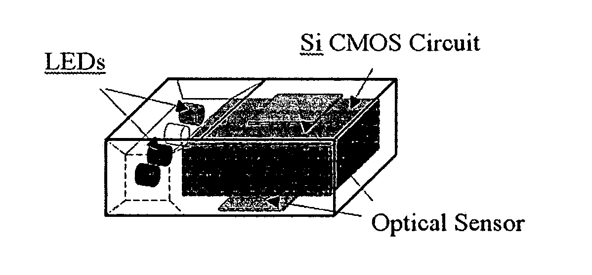
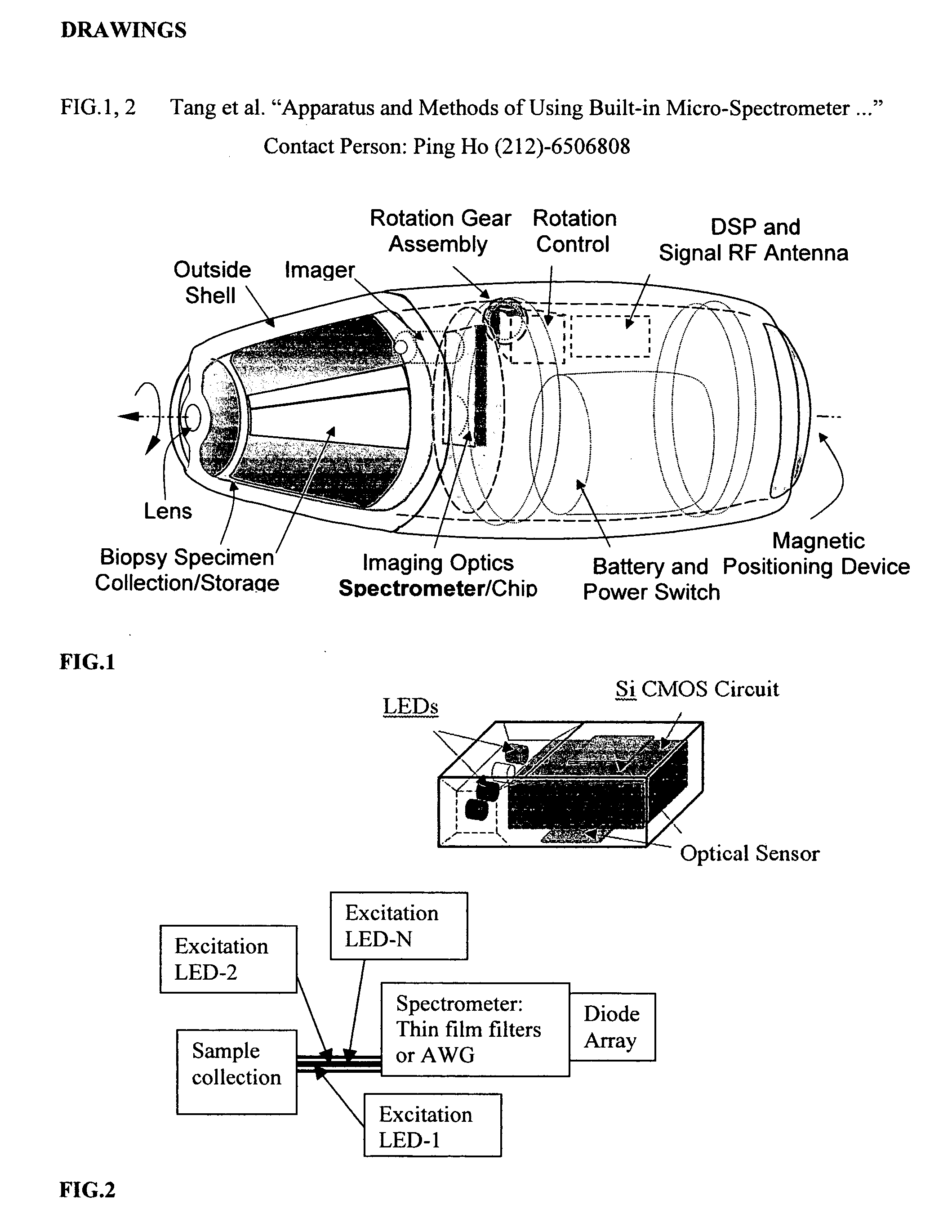
[0059] The principles and preferred embodiments of the present invention is a wireless capsule. A schematic diagram of a wireless capsule for spectroscopic biopsy is shown in FIG. 1. This capsule can travel into a nature tract of a living biological body, e.g., human body by a non-invasive or a minimally invasive procedure such as gastrointestinal (GI) by mouth, and urinary system, biliary tract, cardiovascular system by injection. Furthermore, this capsule can travel to a variety of sites inside the body, such as the esophagus, stomach, biliary tract, gallbladder, pancreatic tract, intestines, colon, rectum, urinary tract cardiovascular tract, and so on.

[0060] The wireless capsule adapted for use inside a biological body will be a capsule without a wire connection, but with or without a remote-control system outside the body. It will be 1 mm to 30 mm in length, 1 mm to 15 mm in wide or in diameter and a form as a cylinder or any other form. It comprises of [0061] (a) a sheath of c...


PUM


Abstract

A wireless capsule as a disease diagnosis tool in vivo can be introduced into a biological body by a native and/or artificial open, or endoscope, or an injection. The information obtained from a micro-spectrometer, and/or an imaging system, or a micro-biosensor, all of which are built-in a wireless capsule, can be transmitted to the outside of the biological body for medical diagnoses. In addition, a real-time specimen collection device is integrated with the diagnostic system for the in-depth in vitro analysis

Description

CROSS REFERENCE TO RELATED PATENT APPLICATIONS [0001] The present application claims the benefit under 35 U.S.C. 119(e) of U.S. Provisional Patent Application Ser. No. 60 / 437,022 filed Dec. 31, 2002. REFERENCES CITED U.S. Patent Documents [0002] Alfano, R. et al. “Remote-controllable, micro-scale device for use in in vivo medical diagnosis and / or treatment”, U.S. Pat. No. 6,240,312, May 29, 2001. [0003] Kim, B. et al. “Micro capsule type robot” U.S. patent application No. 20030092964, May 15, 2003. [0004] Kimchy, Y. et al. “Ingestible device” U.S. patent application No. 20030139661, Jul. 24, 2003. [0005] Georgakoudi, I. et al. “System and methods of fluorescence, reflectance and light scattering spectroscopy for measuring tissue characteristics”, U.S. patent application No. 20030013973, Jan. 16, 2003. [0006] Chance, B. “Optical coupler for in vivo examination of biological tissue,” U.S. Pat. No. 5,987,351, Nov. 16, 1999. [0007] Alfano, R. et al. “Technique for examining biological ...

Claims


Application Information

Patent Timeline
IPC IPC(8): A61B1/05A61B5/00A61B10/00A61B19/00
CPCA61B1/00158A61B1/041A61B1/043A61B5/0031A61B5/0071A61B2019/2249A61B5/0084A61B5/1459A61B10/0045A61B10/0233A61B5/0075A61B34/72
Inventor WANG, LEMINGYING, JINPINTANG, JINGLEE, WEILONGHO, PINGPEI
Owner WANG LEMING
Why Eureka
Try Eureka