
Antigen presenting system and methods for activation of T-cells

a technology of t-cells and presenting systems, applied in the field of t-cell activation systems and presenting systems, can solve the problems of inability to specifically activate cytotoxic t-cells, few successful attempts to use cytotoxic t-cells, and the cost of eliminating a viral infection is the accompanying loss of infected cells, so as to achieve greater t-cell activation

Inactive Publication Date: 2005-07-14
THE SCRIPPS RES INST
View PDF1 Cites 10 Cited by
  • Summary
  • Abstract
  • Description
  • Claims
  • Application Information

AI Technical Summary

Benefits of technology

[0022] In one embodiment, it is particularly preferred to have at least two assisting molecules, one being a costimulatory molecule and the other being an adhesion molecule. It has been found that this combination has a synergistic effect, giving even greater T-cell activation than either of the individual molecules combined. It has also been found to be advantageous and preferable to have at least one of the transfected genes under control of an inducible promoter.
[0023] Using the present invention, it is possible to introduce the peptide to the cell while it is producing MHC molecules and allow the peptide to bind the MHC molecules while they are still within the cell. Alternatively, the MHC molecules can be expressed as empty molecules on the cell surface and the peptide introduced to the cells after the molecules are expressed on the cell surface. In this latter procedure, the use of poikilotherm cells is particularly advantageous because empty MHC molecules, those not yet complexed or bound with peptides, are thermolabile.

Problems solved by technology

While various procedures involving the use of antibodies have been applied in those types of diseases, few if any successful attempts using cytotoxic T-cells have been recorded.
However, no procedures have been available to specifically activate cytotoxic T-cells.
Thus, the cost of eliminating a viral infection is the accompanying loss of the infected cells.
While targeting against viral diseases in general may be accomplished in vivo by vaccination with live or attenuated vaccines, no similar success has been achieved with retroviruses or with cancer cells.
Moreover, the vaccine approach has not had the desired efficacy in immunosuppressed patients.
However, this protocol (known as LAK cell therapy) will only allow the expansion of those CD8+ cells which are already activated.
In fact, it has not been documented that this type of therapy activates any cells with the desired specificity.
Thus, the benefits of LAK cell therapy are controversial at best, and the side effects are typically so severe that many studies have been discontinued.
These “empty” molecules are often unable to reach the cell surface, however, as Class I molecules without bound peptide are very thermolabile.
The presentation of Class I MHC molecules bound to peptide alone has generally ineffective in activating CD8+ cells.

Method used


Image

  • Antigen presenting system and methods for activation of T-cells
  • Antigen presenting system and methods for activation of T-cells
  • Antigen presenting system and methods for activation of T-cells

Examples


example 1

Expression of Human Class I MHC Molecules

A. Preparation of pRmHa-3 Expression Vector

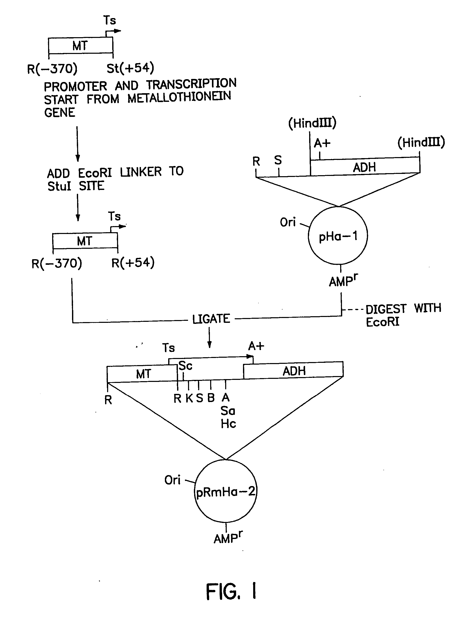
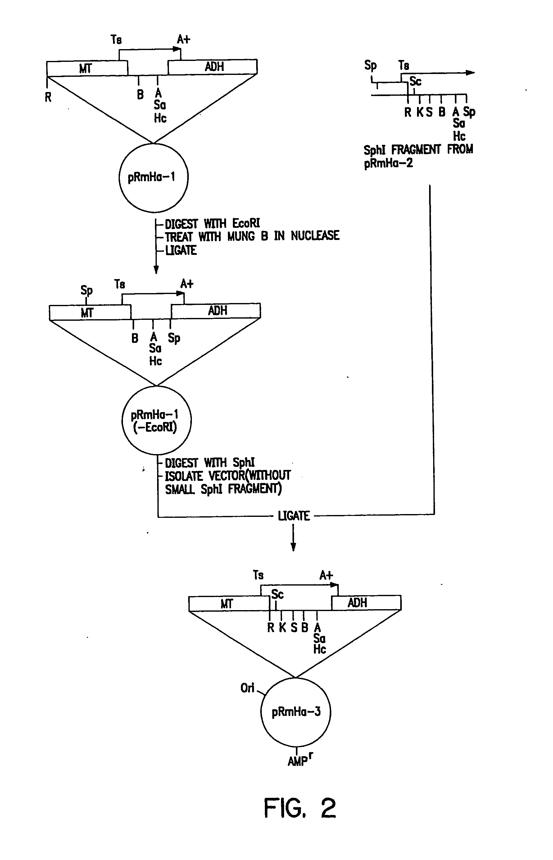
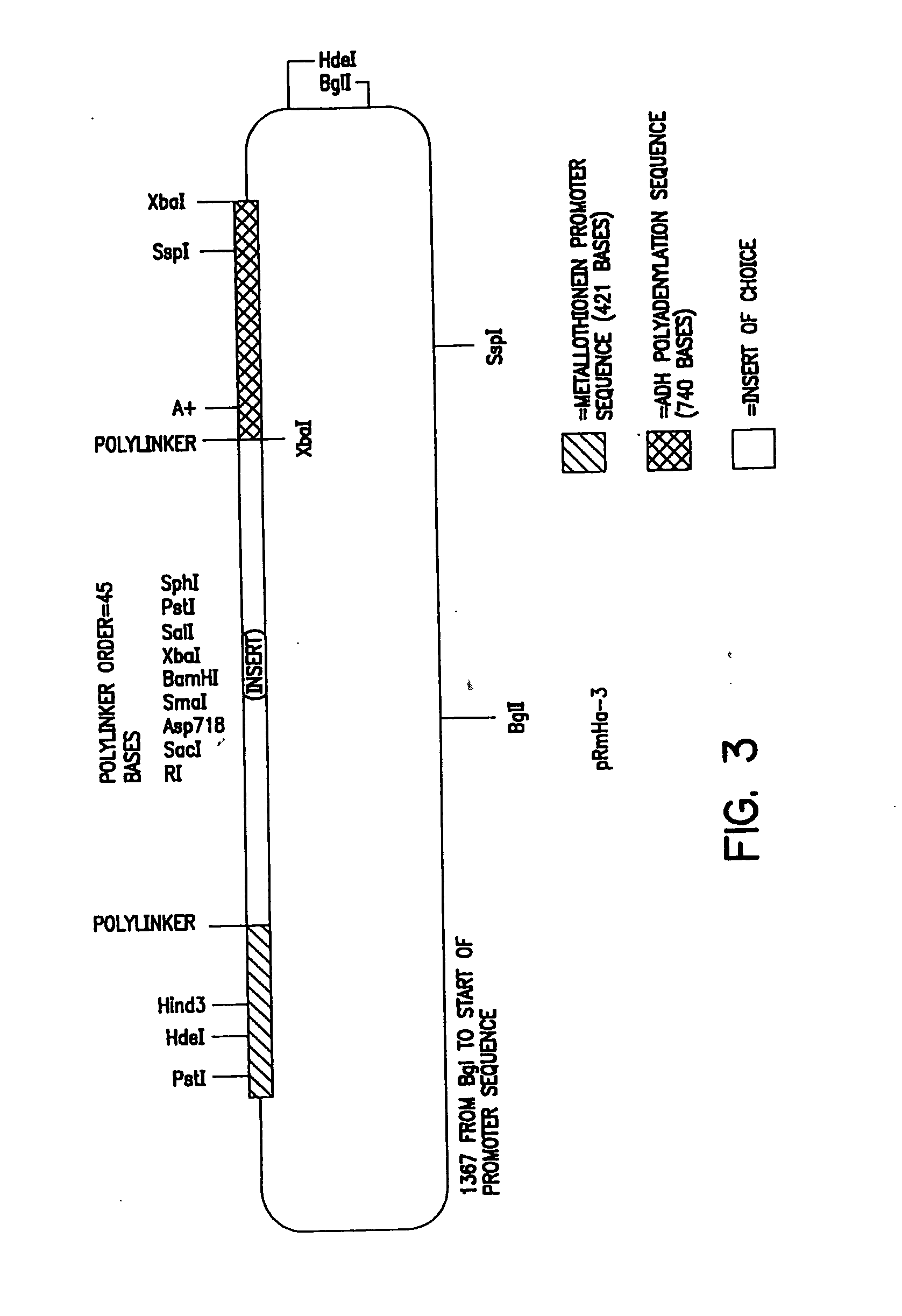
[0150] The pRmHa-3 expression vector for use in expressing MHC proteins in Drosophila Schneider 2 (S2) cells as described in this invention was constructed by ligating a Sph I linearized pRmHa-1 DNA expression vector with a DNA fragment resulting from a Sph I restriction digest of a pRmHa-2 expression vector as described below. The ligating of pRmHa-1 with the pRmHa-2 fragment in this manner was performed to remove one of two Eco RI restriction endonuclease cloning sites present in pRmHa-1. Thus, the resultant pRmHHa-3 expression vector contained only one Eco RI restriction site in the multiple cloning site (polylinker) into which various MHC-encoding DNA fragments were inserted as described in the Examples.

1. Preparation of pRmHa-1 Expression Vector

[0151] The pRmHa-1 expression vector, containing a metallothionein promoter, metal response consensus sequences (designated MT) and an alcohol dehy...

example 2

Preparation of Synthetic Antigen-Presenting Cells

A. Osmotic Loading

[0202] Osmotic loading of SC2 and 3T3 cells with ovalbumin protein was carried out as described by Moore, et al., Cell 54: 777-785 (1988). The assay procedure is as follows. In a 96-well dish, 1×10⁵ Drosophila cells (with or without peptide / protein loaded) or 3T3 cells were cocultured with 1×10⁵ B3 / CD8 T-cell hybridoma cells in 200 μl of RPMI media supplemented with 10% fetal bovine serum. After 24 hours of incubation, 100 μl of the supernatant from these cultures was added to 100 μl of RPMI containing 5,000 CTLL cells. The cells were cocultured for 24 hours at 37° C. when 1 μCi of 3H thymidine (Amersham) was added. After a further incubation of 15 hours at 37° C., the incorporation of radiolabel into the CTLL cells was determined by scintillation counting.

[0203] Assays conducted with murine MHC also verified that the insect cells are capable of loading peptide onto the Class I molecules. Cells expressing as few ...

example 3

Stimulation of Proliferation and Differentiation of Armed Effector T-Cells

[0208] We have found that Drosophila S2 cells transfected with MHC class I molecules and specific assisting molecules are able to stimulate primary responses from T-cells in vitro. We present data below in this example from a mouse model system. In this example, constructs coding for mouse MHC class I (Ld), β2 microglobulin, specific assisting molecules were used and tested with CD8+ cells from lymph nodes of T-cell receptor transgenic mice.

[0209] The data in FIG. 7 provides evidence that the transfected Drosophila S2 cells express the protein products of the transfected murine genes. Flow cytometry using a fluorescence-activated cell sorter (FACS) and fluorescently labeled antibodies were used to demonstrate the expression of Ld (MHC molecule which includes heavy chain and β2) and the specific assisting molecules B7.1 (CD80) and ICAM-1 (CD54) molecules by transfected Drosophila S2 cells. Transfected cells w...


PUM

PropertyMeasurementUnit
temperatureaaaaaaaaaa
Eco RIaaaaaaaaaa

Abstract

The present invention relates to synthetic antigen-presenting matrices, their methods of making and their methods of use. One such matrix is cells that have been transfected to produce MHC antigen-presenting molecules and assisting molecules such as co-stimulatory molecules. The matrices can be used to activate CD8+ T-cells to produce cytokines and become cytotoxic.

Description

CROSS-REFERENCE TO RELATED APPLICATIONS [0001] This application is a division of U.S. patent application Ser. No. 10 / 266,463, filed on Oct. 8, 2002, now U.S. Pat. No. 6,828,150, which, in turn, in a division of U.S. patent application Ser. No. 08 / 913,612, filed on Sep. 8, 1997, now U.S. Pat. No. 6,461,867, which is a national stage application of PCT / US96 / 03249, filed Mar. 8, 1996, which is a continuation-in-part of U.S. patent application Ser. No. 08 / 400,338 filed Mar. 8, 1995, now abandoned.STATEMENT OF GOVERNMENT SUPPORT [0002] This invention was made with the support of the Government of the United States of America under Contract No. CA 38355 by the National Institutes of Health, and the Government of the United States of America has certain rights in the invention.TECHNICAL FIELD [0003] The present invention relates to materials and methods of activating T-cells with specificity for particular antigenic peptides, the use of activated T-cells in vivo for the treatment of a vari...

Claims


Application Information

Patent Timeline
Patent Type & Authority Applications(United States)
IPC IPC(8): C12N15/09A61K35/14A61K38/00A61K39/00A61K48/00A61P35/00C07K14/025C07K14/145C07K14/705C07K14/74C12N5/07C12N5/0783C12N5/10C12N15/12C12N15/85C12P21/02
CPCA61K38/00Y10S530/827C07K14/005C07K14/70503C07K14/70539C12N5/0601C12N5/0636C12N15/85C12N2501/50C12N2501/51C12N2502/50C12N2502/99C12N2760/16122C12N2760/18822C12N2760/20222C12N2830/002C12N2830/75C12N2830/80Y10S530/812A61K2039/5158A61P31/12A61P31/18A61P35/00A61P37/00A61P37/04A61P43/00A61K39/4611A61K39/464838A61K39/464499
Inventor CAI, ZELINGSPRENT, JONATHANBRUNMARK, ANDERSJACKSON, MICHAELPETERSON, PERLUXEMBOURG, ALAINLETURCQ, DIDIERMORIARTY, ANN
Owner THE SCRIPPS RES INST
Learn More