Methods and compositions related to tagging of membrane surface proteins

Inactive Publication Date: 2005-07-21
PROTEOLOGICS INC (US)
View PDF0 Cites 62 Cited by
  • Summary
  • Abstract
  • Description
  • Claims
  • Application Information

AI Technical Summary

Benefits of technology

[0012] In a further embodiment, the invention provides reagents to be used in methods of the invention. Exemplary specific labeling agents are substantially membrane impermeable, and therefore enable selective modification of cell surface proteins. Certain labeling agents of the invention comprise a reversible bond, that facilitates removal o

Problems solved by technology

For example, cancer therapeutics are notorious for their severe side effects, which result largely from a lack of specificity.
Most cancer therapeutics target processes that are common to all growing cells and therefore cause serious damage to healthy cells in addition to cancerous cells.
Membrane-embedded proteins are difficult to ch

Method used


Image

  • Methods and compositions related to tagging of membrane surface proteins
  • Methods and compositions related to tagging of membrane surface proteins
  • Methods and compositions related to tagging of membrane surface proteins

Examples


Example

Example 1

Tagging of Cell Surface Proteins in Live Cells with EZ-LINK NHA-SS-Biotin

[0204] One set of HeLa cells is labeled with cleavable biotin, and a second with DMSO as control. [0205] 1. Wash cells three times with cold PBS. [0206] 2. Detach cells from 4 roller bottles with 50 ml PBS / 5 mM EDTA (prepared at room temperature) for 15 minutes at the incubator, while rolling. Place in a 50 ml tubes and pellet cells at 1800 rpm, 4° C. for 5 minutes. Count cells. [0207] 3. Resuspend cells from all tubes in 50 ml PBS / CM and spin down at 1800 rpm, 4° C. for 10 minutes. [0208] 4. Resuspend the cells at 25×106 cells / ml in PBS / CM containing 0.5 mg / ml sulfo-biotin-NHS. Place cells in a 5 ml snap cup tube and cover with aluminum foil. [0209] 5. Incubate with gentle shaking, in the cold cabinet, for 20 minutes. Spin down cells, 1500 rpm, 4° C. for 5 minutes. Resuspend at 25×106 cells / ml in 0.5 mg / ml PBS / CM containing 0.5 mg / ml sulfo-biotin-NHS. Incubate as before for 20 more minutes. [0210] 6...

Example

Example 2

Tagging of Cell Surface Proteins in Live Cell with Alexa Fluor®488-NHS

[0221] There are several products of Alexa Fluor, each has a different emission maximum. Thus, cells can be treated differently and labeled with different emitting Alexa Fluor reagents. The detection can be done with two lasers, one that will detect one fluorophore and the other the second. An image can then be generated and the proteins that are found in both samples will give a different color then each flourphore alone.

[0222] One set of HeLa cells is labeled with Alexa Fluor®488-NHS, and a second with DMSO as control. All steps are performed on ice to prevent internalization of cell surface proteins. [0223] 1. Wash cells three times with cold PBS. [0224] 2. Detach cells from 4 roller bottles with 50 ml PBS / 5 mM EDTA (prepared at room temperature) for 15 minutes at the incubator, while rolling. Place in a 50 ml tubes and pellet cells at 1800 rpm, 4° C. for 5 minutes. Count cells. [0225] 3. Resuspend ...

Example

Example 3

Cell Surface Protein Profiling Methodologies

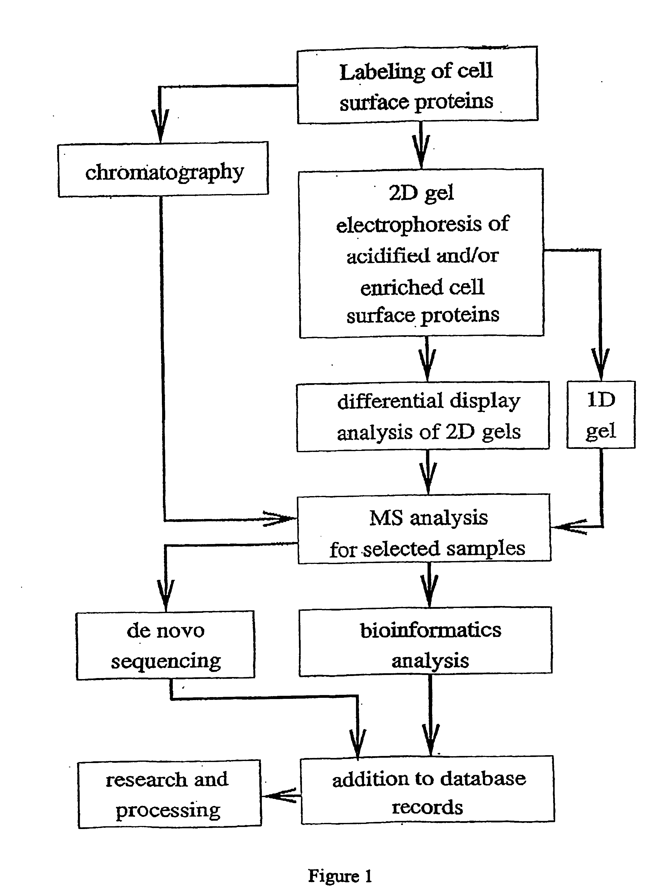
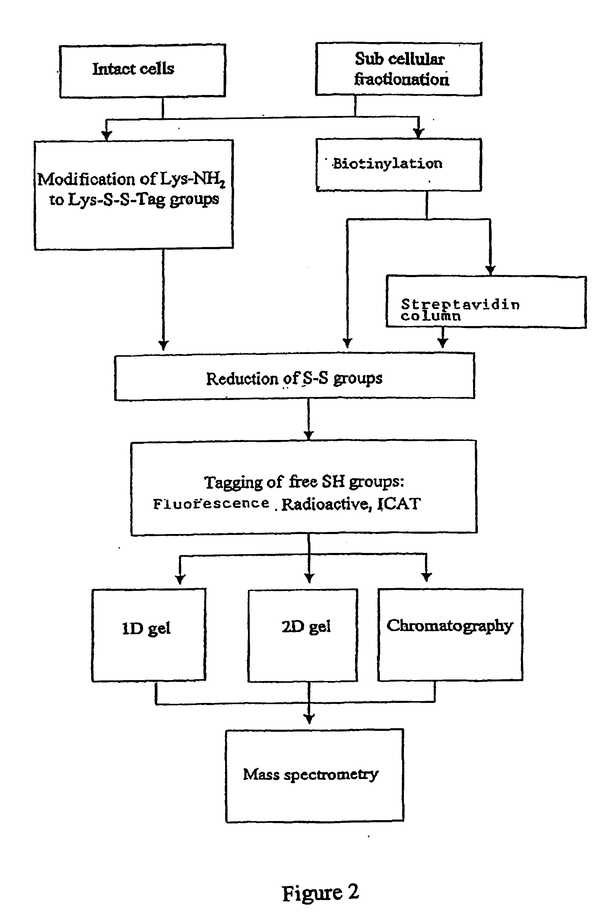
[0237] The flow chart in FIG. 1 exemplifies several possible combinations of cell surface protein labeling and identification techniques. A summary of certain aspects of the illustrated methods is set forth below.

[0238] These exemplary methods begin with a selective labeling of cell surface proteins. The labeling method, when performed using a labeling agent that binds to lysine residues, acidifies proteins, making isoelectric focusing (and thereby 2D gel electrophoresis) possible for highly basic proteins. The labeled proteins are ultimately identified by mass spectrometry analysis. Resolution of proteins for mass spectrometry may be accomplished by chromatographic separations, 2D gel electrophoresis or 1D gel electrophoresis. 2D gel electrophoresis may also be used as a part of differential display method for identifying those proteins whose expression levels change in different conditions.

[0239] MS analysis provides a wealt...


PUM

PropertyMeasurementUnit
Compositionaaaaaaaaaa
Structureaaaaaaaaaa
Surfaceaaaaaaaaaa

Abstract

This invention relates to methods and reagents for selectively labeling membrane surface proteins using a labeling agent. The label may be used to isolate preparations of membrane surface proteins. Preparations of membrane surface proteins may be analysed by a variety of high-throughput techniques to allow rapid profiling of membrane surface protein composition.

Description

CROSS-REFERENCE TO RELATED APPLICATIONS [0001] This application claims priority to U.S. Provisional Application No. 60 / 296,334, filed Jun. 6, 2001 and incorporated by reference herein in its entirety.BACKGROUND [0002] Proteins associated with the plasma membrane constitute a significant and functionally important fraction of the proteins in a cell. Key functions, such as the communication of a cell with its environment, are largely dependent on membrane proteins. Membrane proteins are targets of choice for pharmaceuticals in part because of their exposure to the extracellular environment. Furthermore, cell surface proteins are excellent markers for use in cell sorting and identification because cells need not be damaged in order to detect these proteins. [0003] The clinical importance of membrane proteins may be illustrated through an examination of the diagnosis and treatment of various cancers. For example, cancer therapeutics are notorious for their severe side effects, which res...

Claims


Application Information

Patent Timeline
IPC IPC(8): C07D207/40C07D207/416C07D319/06C07D405/12C07D491/04C07D491/14C07F5/02C07K1/107C07K1/13C07K14/705C12Q1/70G01N33/50G01N33/53G01N33/554G01N33/567G01N33/58G01N33/68
CPCC07D207/416C07D319/06C07D405/12C07D491/04C07D491/14C07F5/025G01N33/6803C07K1/13C07K14/705G01N33/5005G01N33/554G01N33/582C07K1/1077
Inventor ALROY, IRISMOSKOWITZ, HAIMREISS, YUVALSHOHAM, BENJAMIN A.
Owner PROTEOLOGICS INC (US)
Why Eureka
Try Eureka