Registration of ultrasound to fluoroscopy for real time optimization of radiation implant procedures

a radiation implant and fluoroscopy technology, applied in the field of ultrasound and carm fluoroscopy imagery registration, can solve the problems of not being able to meet the most likely conditions of c-arms, few attempts to relate fluoroscopic images to soft tissue anatomy with little success, and not being able to achieve quantitative intra-operative analysis, etc., to achieve the effect of improving the localization of radiation doses, improving visualization and control, and avoiding the use of c-arms

Inactive Publication Date: 2005-08-04
THE JOHN HOPKINS UNIV SCHOOL OF MEDICINE
View PDF10 Cites 38 Cited by
  • Summary
  • Abstract
  • Description
  • Claims
  • Application Information

AI Technical Summary

Benefits of technology

[0013] An advantage of the present invention is to provide improved visualization and control during radiation implant processes.
[0014] Another advantage of the present invention is improved localization of radiation doses in cancer treatment.
[0015] Another advantage of the present invention is to enable an advantageous compromise between time, dose, and accuracy during radiation dose implant surgery.
[0016] Another advantage of the present invention is to better enable dynamic dose calculation during radiation implant procedures, including the ability to adjust the planned implant locations to better optimize dose as the implant progresses.

Problems solved by technology

While C-arm fluoroscopy has been used intra-operatively, it has not been utilized in quantitative intra-operative analysis.
Very few attempts have been made to relate fluoroscopic images to soft tissue anatomy with little success.
Here the fundamental problem is matching large number of seeds with their projections in multiple X-ray images when some seeds obscure each other and solid objects also can get in the way.
Automated methods have been explored, but they also assume conditions that most likely cannot be met on a C-arm, particularly in a realistic intra-operative scenario.
Such assumptions include no extrinsic object in the field, optimal beam energy, arbitrary number and orientation of X-ray shots, or unlimited processing time and computational resources—none of which is realistic in real-time image-guided surgery.
In conclusion, “off-the-shelf” post-implant seed matching and reconstruction techniques cannot be expected to work in the operating room.
Many approaches have met with great difficulty due to the inability to accurately determine the imaging angles relative to the prostate for reconstruction of the multiple projection images.
This not only reduces accuracy, but makes the process too lengthy to be used intraoperatively.
The use of implanted needles as fiducial markers for registration of biplane TRUS data has been explored, but several key problems have been left unsolved: (1) The use of implanted needles as fiducials may not be practical, because most practitioners implant only one needle at a time and they do not use stabilizing needles.
During TRUS scanning, however, the prostate deforms, dislocates, and the needles dislocate relative to the prostate.
(4) Previous attempts did not account for the need to pre-operatively calibrate the C-arm fluoroscope, including removing image distortion and some form of intra-operative tracking to know where the multiple X-ray shots are coming with respect to one another.
Currently known dewarping and calibration kits are custom designed for each C-arm, making them hard to transfer from device to another.
Ultrasound has been an excellent tool in guiding the implant needles with respect to prostate anatomy, yet it cannot show reliably the location of radioactive seeds after they are released in the prostate.
Intraoperative C-arm fluoroscopy can show the implanted seeds, but it cannot detect prostate anatomy.

Method used


Image

  • Registration of ultrasound to fluoroscopy for real time optimization of radiation implant procedures
  • Registration of ultrasound to fluoroscopy for real time optimization of radiation implant procedures
  • Registration of ultrasound to fluoroscopy for real time optimization of radiation implant procedures

Examples


Embodiment Construction

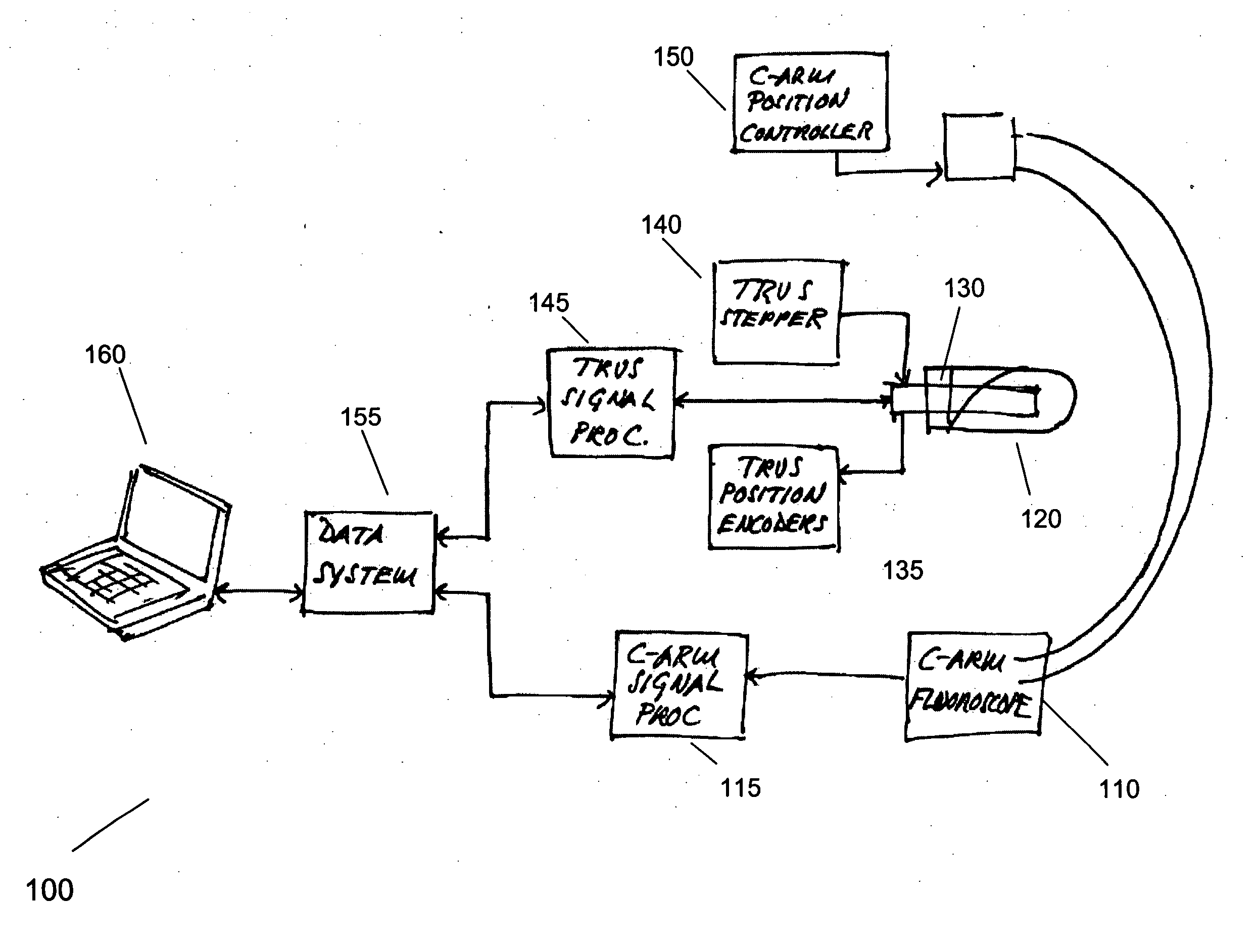
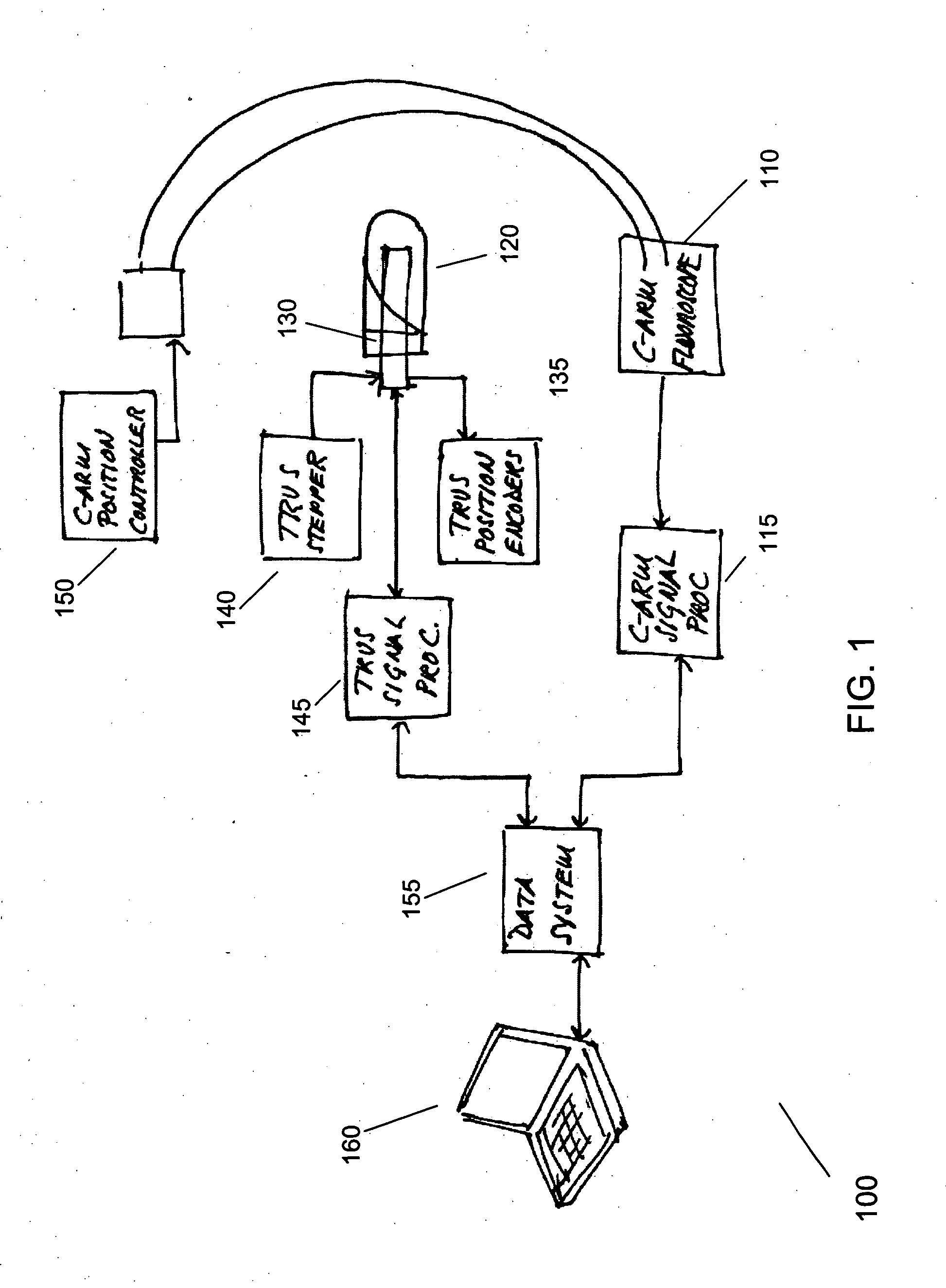
[0030]FIG. 1 shows an exemplary system 100 for registering ultrasound and C-arm fluoroscopy according to the present invention. The system 100 generally comprises commercially available TRUS and C-arm fluoroscopy hardware. The system 100 includes a C-arm fluoroscope 110; related C-arm fluoroscope signal processing hardware and software 115; a C-arm position controller 150; a transrectal sheath 120; a TRUS probe 130; a TRUS stepper; a TRUS probe position encoder 135; TRUS signal processing hardware and software 145; a data system software 155; and a computer 160, which stores and executes the data system software 155. The computer 160 may comprise multiple computers, including remote databases and embedded processors. It will be apparent to one skilled in the art that the data system software 155, and the computer 160, may be provided in many different configurations.

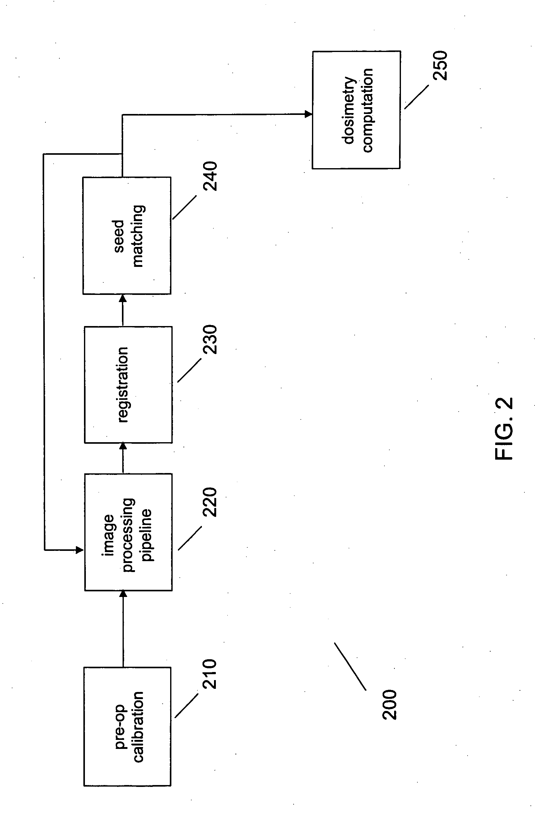
[0031]FIG. 2 shows an exemplary process 200 for registering ultrasound and C-arm fluoroscopy according to the present...


PUM


Abstract

Transrectal ultrasound guided transperineal low dose-rate brachytherapy has been emerged as one of the definitive treatments of low-risk prostate cancer. Ultrasound has been an excellent tool in guiding the implant needles with respect to prostate anatomy, yet it cannot show reliably the location of radioactive seeds after they are released in the prostate. Intraoperative C-arm fluoroscopy can show the implanted seeds, but it cannot detect prostate anatomy. Intra-operative fusion of these two complementary modalities offers significant clinical benefit by allowing for real-time optimization of the brachytherapy implant as the procedure progresses in the operating room. Disclosed is a system and method for mitigating this problem and providing registration of seeds seen by fluoroscopy with live prostate anatomy visualized by transrectal ultrasound.

Description

[0001] This application claims the benefit of U.S. Provisional Patent Application No. 60 / 488,965, filed on Jul. 21, 2003, which is hereby incorporated by reference for all purposes as if fully set forth herein.[0002] Research and development efforts associated with the subject matter of this patent application was supported by the National Science Foundation under Grant No. #ERC 9731478.BACKGROUND OF THE INVENTION FIELD OF THE INVENTION [0003] The present invention relates to the registration of ultrasound and C-arm fluoroscopy imagery for the purposes of providing real-time optimization of medical procedures such as transperineal low-dose rate brachytherapy. [0004] Adenocarcinoma of the prostate is the most commonly diagnosed cancer in the U.S. male population. During the last half decade, there have been approximately 200,000 new cases of prostate cancer diagnosed each year, which is comparable to breast cancer diagnosis, and there is no evidence that this number would significant...

Claims


Application Information

Patent Timeline
Patent Type & Authority Applications(United States)
IPC IPC(8): A61BA61B5/05A61B6/00A61B8/00A61B8/12A61B17/00A61N5/00G21K4/00
CPCA61B6/504A61B8/12A61B6/583A61B6/5247A61B6/4417A61B2090/392A61B90/39A61B2090/3925A61B2090/3937A61B2090/3954A61B2090/3966A61N5/1014
Inventor FICHTINGER, GABORMUSTUFA, TABISHWYROBEK, KEENANBURDETTE, E. CLIF
Owner THE JOHN HOPKINS UNIV SCHOOL OF MEDICINE
Why Eureka
Try Eureka