Micro-scale compact device for in vivo medical diagnosis combining optical imaging and point fluorescence spectroscopy

a compact device and micro-scale technology, applied in the field of medical imaging apparatus for biological tissue visualization and assessment, can solve the problems of high cost and harmful radiation exposure, time consumption of x-ray imaging, and the inability of mri and ct scanning to provide detailed photographic images of the surface of sampled tissue when desired, so as to achieve accurate tissue detection and time-consuming, non-invasive

Active Publication Date: 2005-09-29
THE CITY COLLEGE OF THE UNIV OF NEW YORK
View PDF22 Cites 98 Cited by
  • Summary
  • Abstract
  • Description
  • Claims
  • Application Information

AI Technical Summary

Benefits of technology

[0012] The present invention is directed to improving the aforesaid problems which occur in the prior art and to provide an apparatus and a method for the timely, non-invasive and accurate detection of tissue and cell normalities and abnormalities, including pre-cancer, cancer arteriosclerosis, and the like.

Problems solved by technology

Disadvantages of X-Ray imaging, MRI and CT scanning include time consumption, high cost and harmful radiation exposure.
Moreover X-Ray imaging, MRI and CT scanning cannot provide detailed photographic images of surfaces of sampled tissue when desired.
Moreover, as tissue samples are usually only taken at sites which have visual evidence of a malignancy, the detection of pre-cancerous, or early stage cancerous tissue is delayed, which can have catastrophic results.
Currently, endoscopy is an invasive method of imaging which requires that the patient be anesthetized and which can cause discomfort or pain to the patient after the procedure.
Moreover, endoscopy is expensive and time consuming, and cannot immediately confirm the presence of cancer cells or pre-cancerous cells.
Furthermore, the removal of tissue specimens for biopsies can cause additional discomfort or pain to the patient after the procedure.
Although the use of a point source fluorescence for the detection of cancer has been demonstrated in areas of the body which do not require the use of a endoscope, such as areas of the gynecological (GYN) tract, aerodigestive tract, colon, breast and esophagus, in vivo fluorescence imaging through an endoscope has presented formidable problems such as: currently available imaging endoscopes use imaging fibers manufactured from glass and therefore cannot transmit the required UV and blue light; detectors located on the distal end of endoscopes have poor UV spectral response; and fluorescence signals in UV and blue light are weaker than scattered light and thus require electronic amplification.
Unfortunately, the use of fluorescence spectroscopy and imaging in a single micro-scaled device capable of both visually imaging and assessing tissue needs improvement.

Method used


Image

  • Micro-scale compact device for in vivo medical diagnosis combining optical imaging and point fluorescence spectroscopy
  • Micro-scale compact device for in vivo medical diagnosis combining optical imaging and point fluorescence spectroscopy
  • Micro-scale compact device for in vivo medical diagnosis combining optical imaging and point fluorescence spectroscopy

Examples


Embodiment Construction

[0034] A preferred embodiment of the present invention will now be described in more detail.

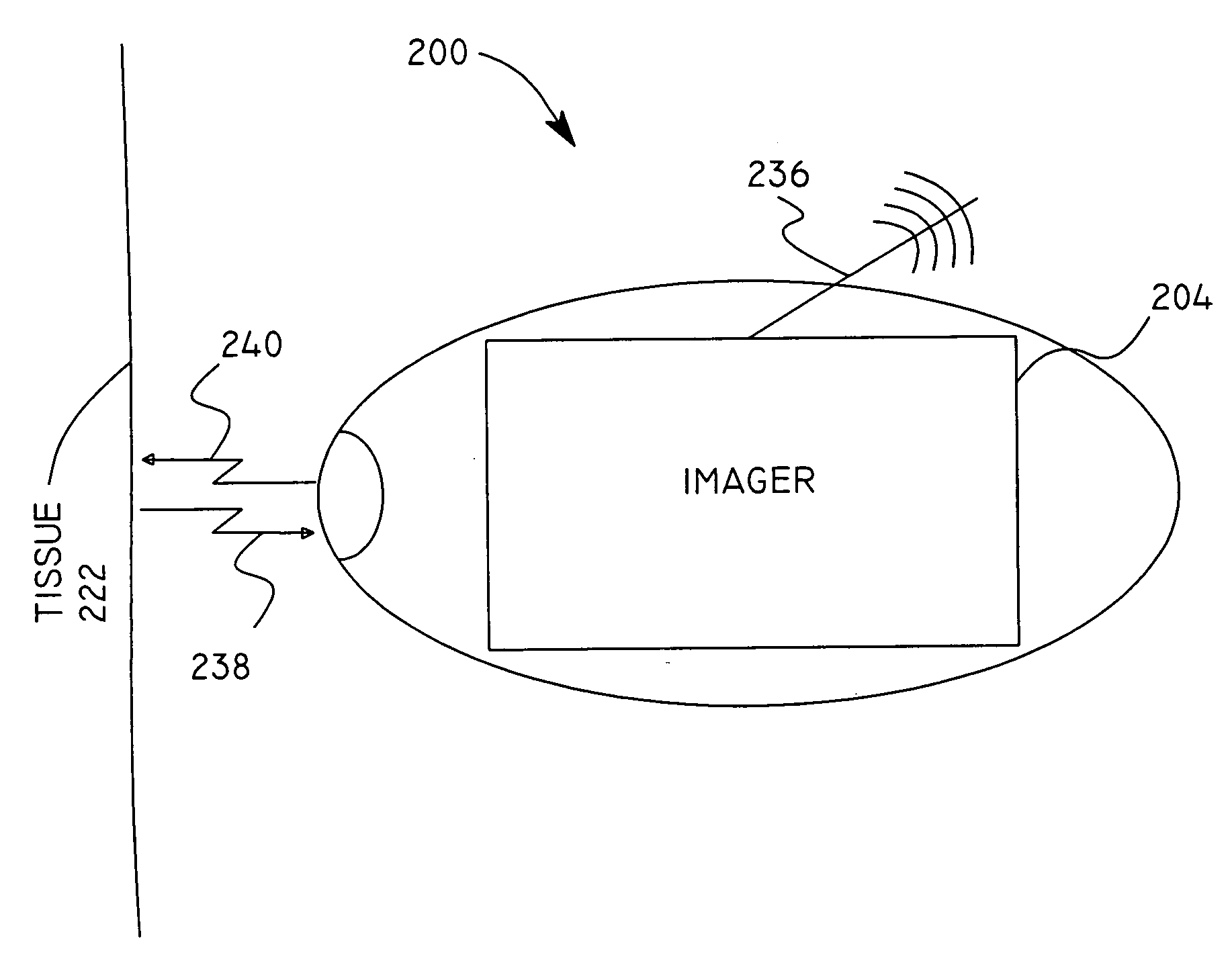
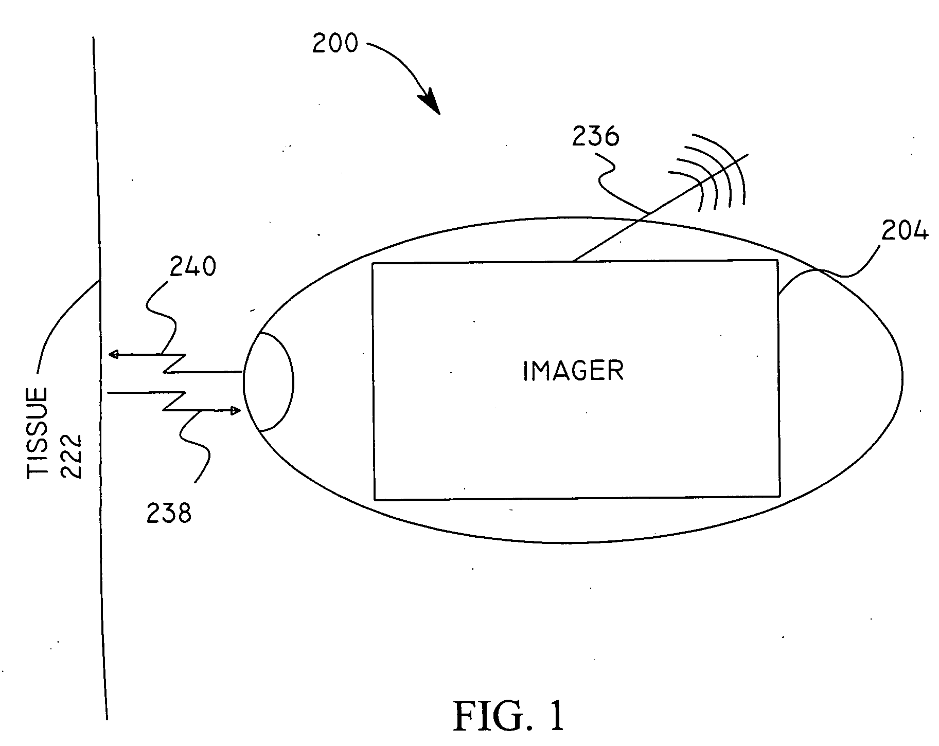
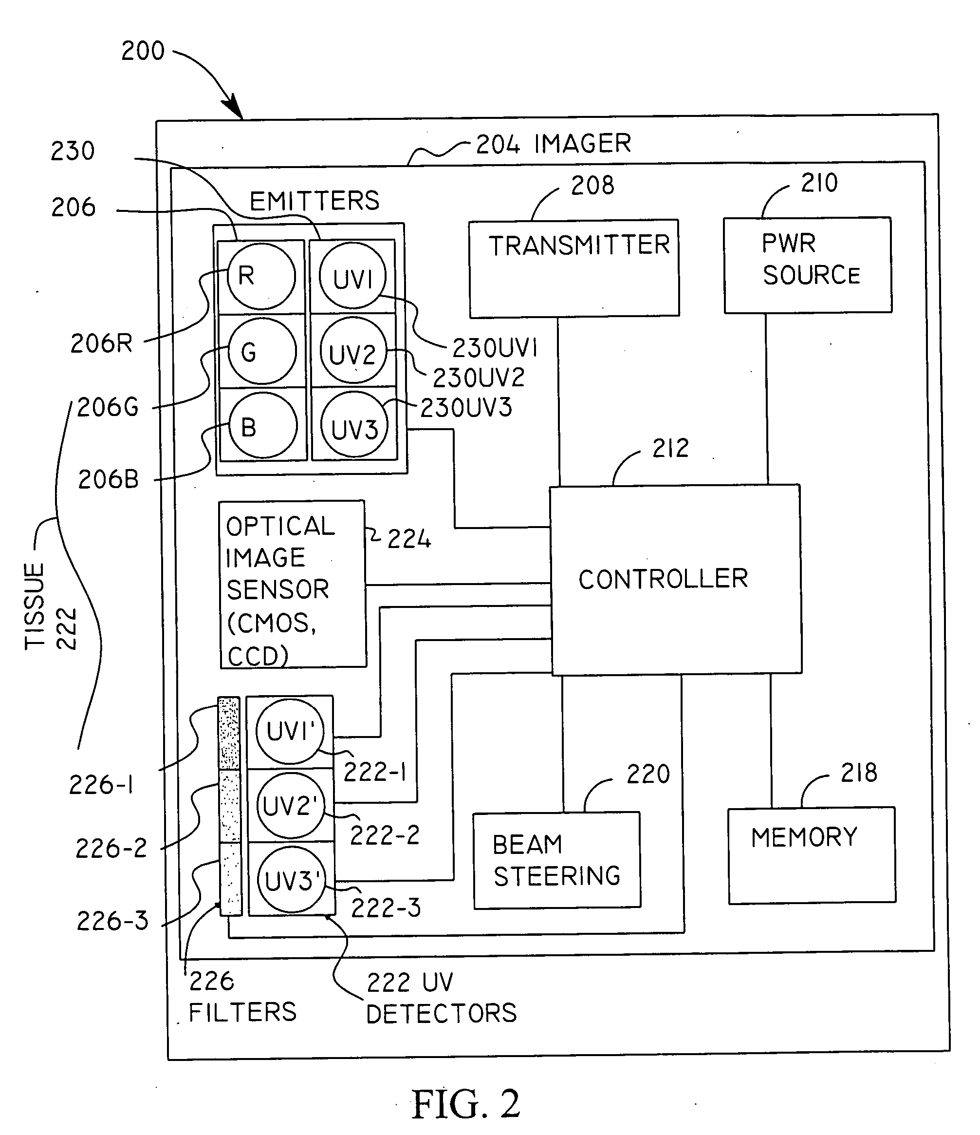
[0035] The present invention is directed to the use of a medical imaging and assessment apparatus and method including CMOS or CCD sensors, UV light sources, UV / visible point sensors, visible light sources, at least one visible imaging detector, dispersion selection components, and beam steering components (e.g., lenses, gratings, mirrors, prisms, optical fibers, beam-splitters, combinations of these, or like) to image an area of tissue combined with point fluorescence spectroscopy of the area in order to image and detect and distinguish normal areas from abnormal areas (e.g., areas having pre-cancerous and cancerous cells and the like). Moreover, the present invention provides data (e.g., image and fluorescence information including fluorescence intensity) to the operator (e.g., a surgeon, or other medical provider) in real time for real-time diagnosis and detection of abnormalities such as...


PUM


Abstract

An apparatus and method for medical practitioners to detect the presence of abnormal cells including cancerous and pre-cancerous cells by using a transport capsule containing an imaging apparatus including UV sources and fluorescence detectors for obtaining images and fluorescence data of biological cells and tissue. The method includes the steps of scanning biological tissue using an ultra-violet (UV) source to obtain fluorescence data, transferring fluorescence data and/or images using a radio frequency (RF) or other suitable means to a personal computer (PC) system, analyzing the image and/or fluorescence data in the PC, identifying -tissues with precancerous and cancerous cells, and optionally determining their precise location, and assessing the accuracy of the calculated fluoroscopic images.

Description

PRIORITY [0001] This application claims priority to a provisional patent application entitled “Micro-Scale Compact Device For In Vivo Medical Diagnosis Combining Optical Imaging And Point Fluorescence Spectroscopy”, filed in the United States Patent and Trademark Office on Jan. 16, 2004 and assigned Ser. No. 60 / 536,723, the contents of which are hereby incorporated by reference.BACKGROUND OF THE INVENTION [0002] 1. Field of the Invention [0003] The present invention relates generally to a medical imaging apparatus for biological tissue visualization and assessment and more particularly to a medical imaging apparatus and method for the detection of abnormal biological tissue including cancerous and precancerous cellular regions of arteries for detection of arterial disease. [0004] 2. Description of the Related Art [0005] With the pervasiveness of cancer in modem times, it has become common to screen the aeorodigestive tract, gynecological (GYN) tract and breast(s) for malignancies. T...

Claims


Application Information

Patent Timeline
Patent Type & Authority Applications(United States)
IPC IPC(8): A61B1/04A61B5/00A61B6/00
CPCA61B1/041A61B5/0084A61B5/0071A61B1/043A61B1/0638A61B1/0684
Inventor ALFANO, R. R.KATZ, ALVINALFANO, SCOTT
Owner THE CITY COLLEGE OF THE UNIV OF NEW YORK
Why Eureka
Try Eureka