Detection of nitrosylated proteins

a technology of nitrosylated proteins and detection methods, applied in the direction of electrolysis, isotope separation, enzyme stabilisation, etc., can solve the problem of limitations in the methodology typically used

Inactive Publication Date: 2005-10-27
UNIVERSITY OF VERMONT
View PDF5 Cites 7 Cited by
  • Summary
  • Abstract
  • Description
  • Claims
  • Application Information

AI Technical Summary

Benefits of technology

[0007] The present invention generally relates to systems and methods for determining oxidized proteins, such as nitrosylated proteins. In some cases, the present invention relates to visualization techniques for spatially determining the spatial locations and / or concentrations of nitrosylated or otherwise oxidized proteins within cells and / or tissues. The subject matter of the present invention involves, in some cases, interrelated products, alternative solutions to a particular problem, and / or a plurality of different uses of one or more systems and / or articles.

Problems solved by technology

This is primarily due to limitations in the methodology typically used to detect S-nitrosylated proteins.

Method used


Image

  • Detection of nitrosylated proteins
  • Detection of nitrosylated proteins
  • Detection of nitrosylated proteins

Examples


example 1

[0047] This example describes certain protocols and methods that may be useful in various embodiments of the invention.

[0048] A line of spontaneously transformed mouse lung alveolar type II epithelial cells (C10) was used in some experiments, as described below. C10 cells were cultured in CMRL1066 medium containing 10% fetal bovine serum (FBS, GIBCO). In experiments involving microscopic analysis, the cells were grown on glass cover slips. At about one hour prior to exposure to a test agent, the medium was switched to phenol red free DMEM / F12 containing 0.5% FBS. Some cells were exposed to S-nitroso-glutathione (GSNO) or S-nitroso-N-acetyl penicillamine (SNAP) for about 1 hour, in the presence of an equimolar concentration of freshly prepared L-cysteine. To monitor S-nitrosylation in response to NOS2 activation, the cells were treated with 10 ng / ml TNF alpha (TNFα) and 100 U / ml interferon gamma (IFNγ), for about 24 hours. Incubation with L-N-monomethyl arginine (L-NMMA) for about 1...

example 2

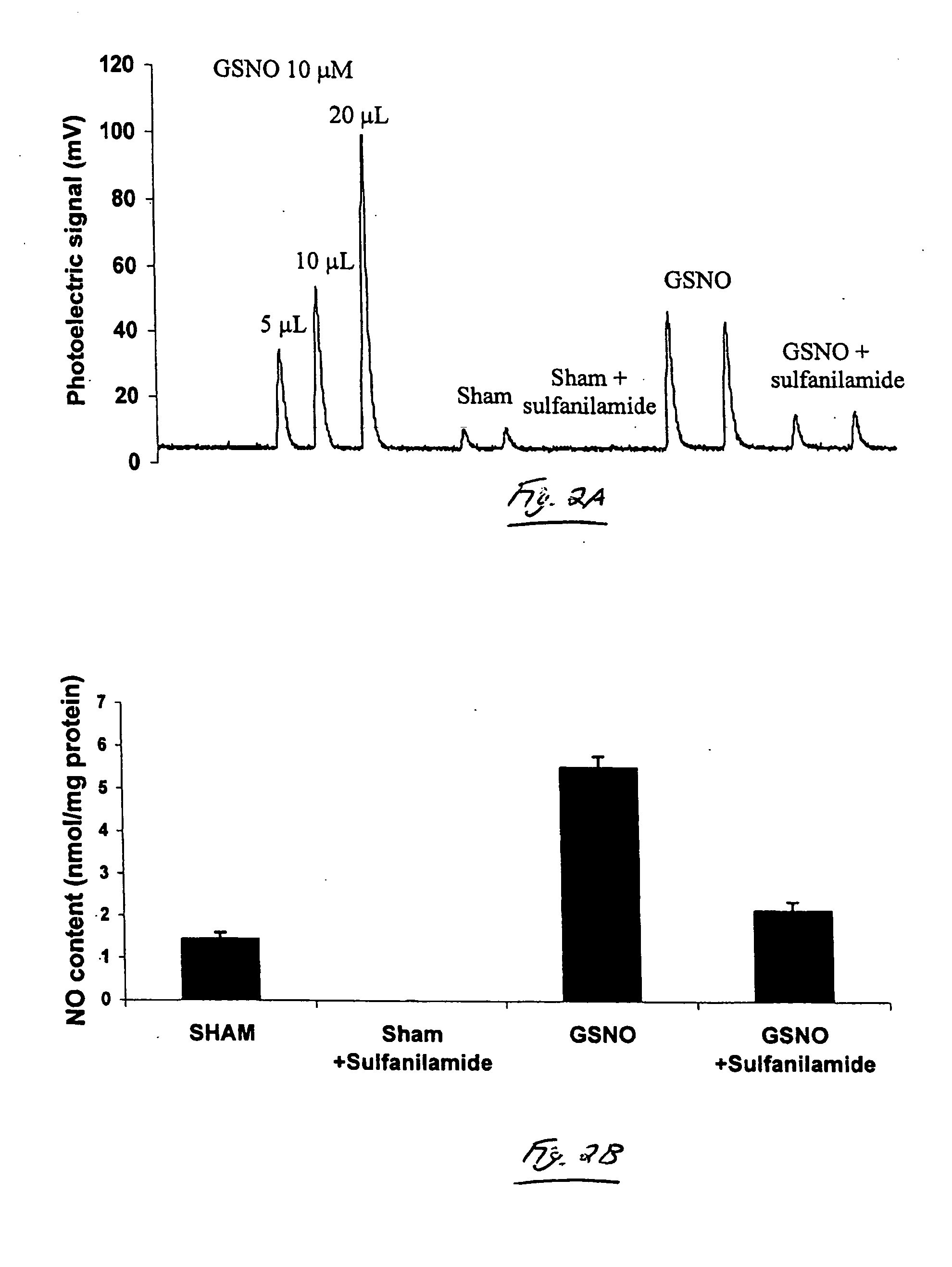
[0054] This example demonstrates NO detection by chemiluminescence, using one embodiment of the invention. Using methods similar to those described in Example 1, this example demonstrates that the treatment of cells with exogenous S-nitrosothiols resulted in an increase in the SNO (nitrosothiol) content of the cells.

[0055] Mouse lung epithelial (C10) cells were incubated with 1 mM GSNO for about one hour, lysed, and the NO released following reduction in acidic KI / I2 was detected by chemiluminescence. As shown in FIG. 2, the total NO content in lysates from cells treated with GSNO was found to have increased, as compared with lysates from non-treated cells. In order to eliminate the contribution of nitrites to the observed photoelectric signal, the cell lysates were incubated with a nitrite quencher, sulfanilamide, and assessed the sulfanilamide resistant NO signal by chemiluminescence. Under these experimental conditions, employing 12 micrograms of protein, S-nitrosthiol was not d...

example 3

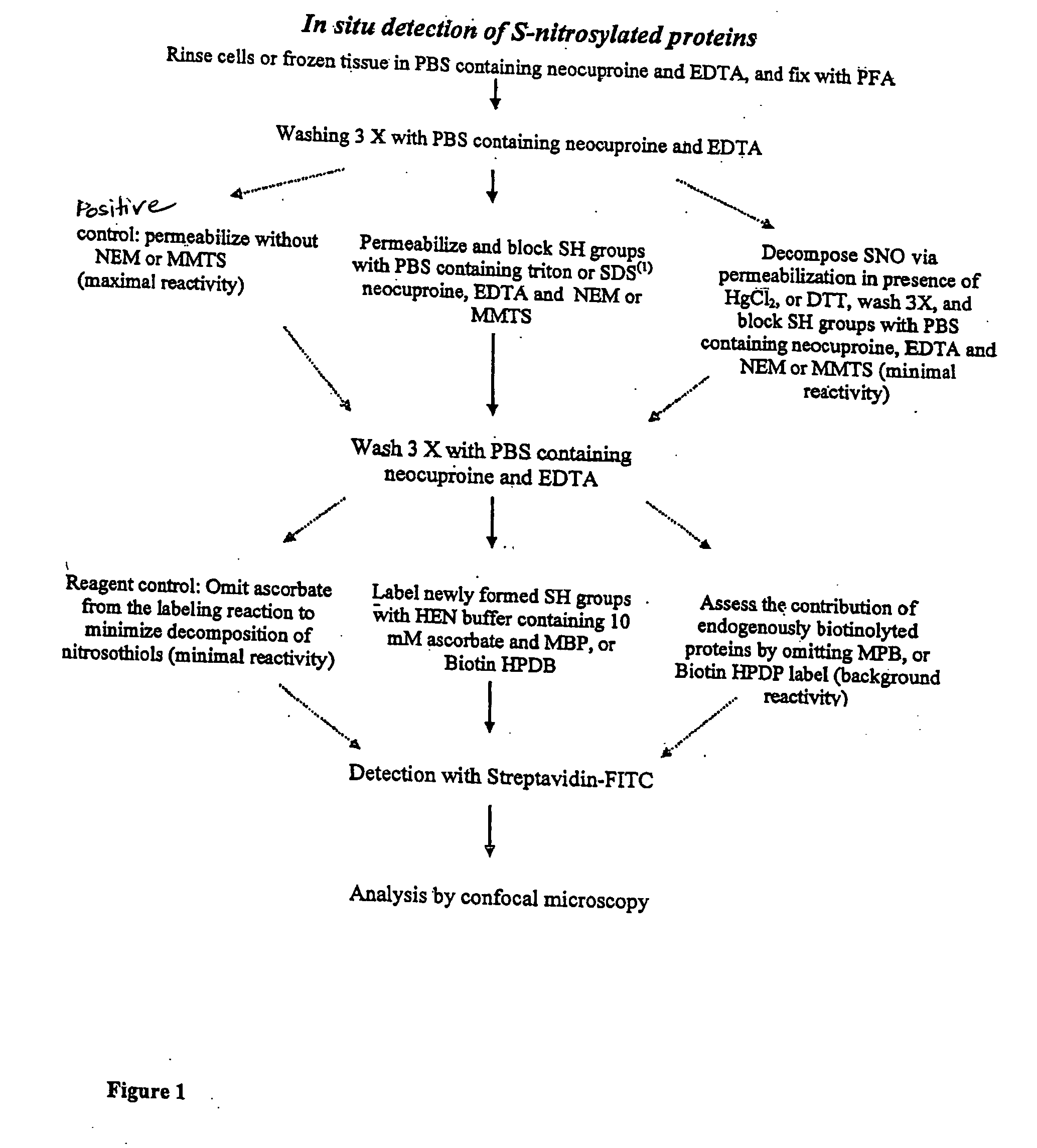
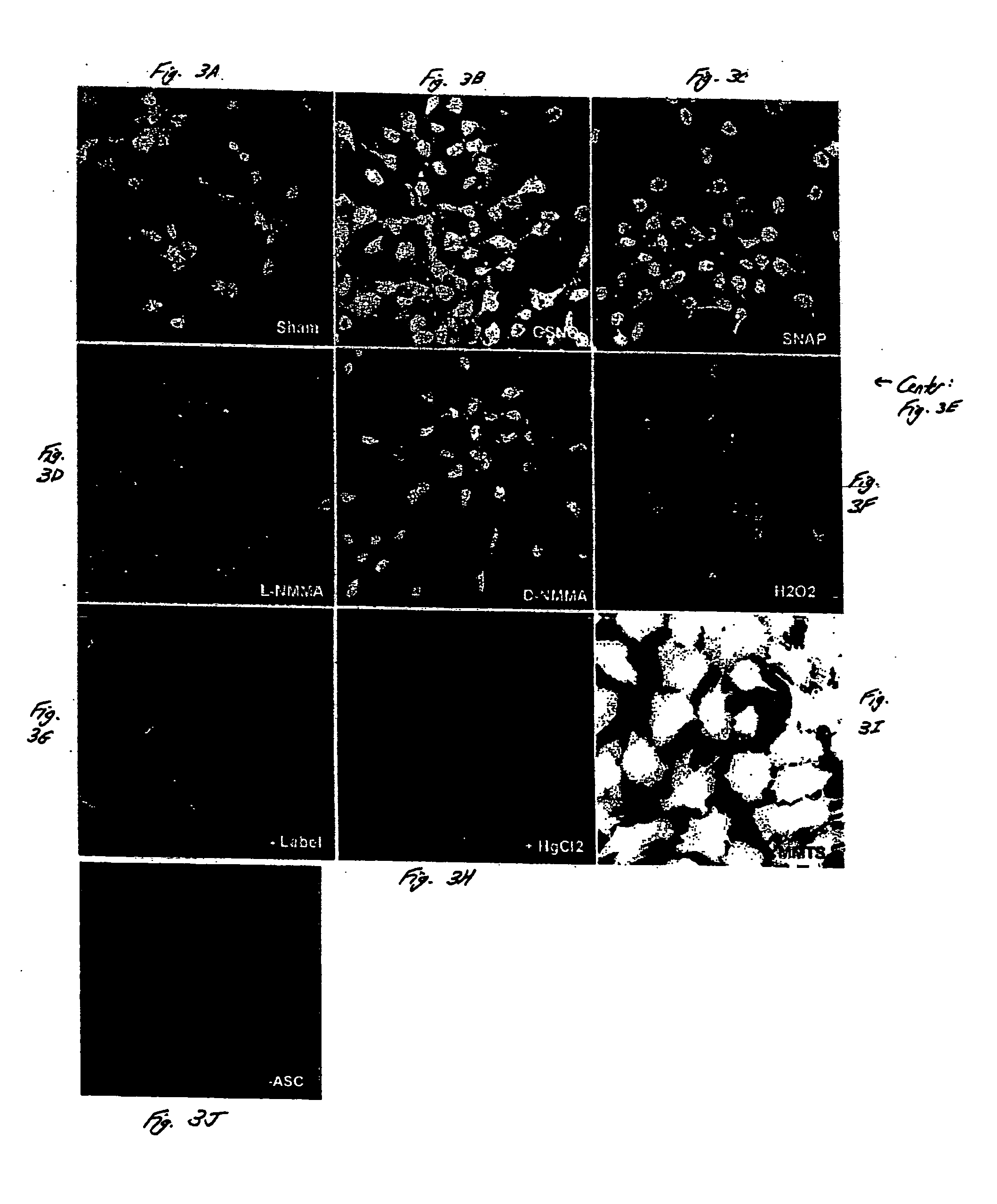
[0057] The detection of S-nitrosylated proteins in situ is demonstrated in this example, in accordance with an embodiment of the invention. Intact C10 cells were studied using methods similar to those described in Example 1. As shown in FIG. 3, S-nitrosylated proteins were detected in intact C10 cells using biotinylation, streptavidin-FITC and analysis by confocal laser scanning microscopy. C10 cells were exposed to 1 mM SNAP (FIG. 3C) or 1 mM GSNO (FIG. 3B) for about 1 hour, or to L-NMMA (FIG. 3D) or D-NMMA (FIG. 3E) for about 16 hours, fixed, and subjected to biotin derivatization, incubation with streptavidin-FITC, and confocal microscopy.

[0058] In non-treated cells (sham controls, FIG. 3A), some endogenous reactivity was observed. This reactivity was inhibited upon the addition of HgCl2, which decomposes SNO prior to blocking with methyl methanethiosulfonate (MMTS) (FIG. 3H). The omission of ascorbate, which reduces SNO bonds prior to the addition of the biotin-HPDP label, also...


PUM

PropertyMeasurementUnit
pHaaaaaaaaaa
pHaaaaaaaaaa
temperatureaaaaaaaaaa

Abstract

The present invention, in some aspects, relates to systems and methods for determining oxidized proteins, including nitrosylated proteins such as S-nitrosylated proteins. The systems and methods of the invention can be used in vitro (e.g., in cell or tissue culture) or in vivo. For instance, in some cases, the invention can be used to spatially determine the location and/or concentration of oxidized proteins within cells and/or tissues (e.g., through visual detection). In one set of embodiments, a nitrosylated or otherwise oxidized moiety on a protein may be reacted with a detection entity, which may be, for example, fluorescent, radioactive, electron-dense, able to bind to a signaling entity or a binding partner in order to produce a signal, etc. As a specific example, a nitrosothiol moiety on a nitrosylated protein may be reacted with an alkylating agent to form an alkylthio moiety; the alkylthio moiety may include a detection entity or otherwise be able to interact with a signaling entity. In some embodiments, other moieties on the protein may be altered or blocked before reaction of the protein with the detection entity. Such moieties on the protein may be, for instance, non-oxidized or non-nitrosylated moieties able to react with the detection entity. As a particular example, in a protein containing nitrosothiol and (non-nitrosylated) thiol moieties, the thiol moieties may first be altered or blocked prior to reaction of the protein with the detection entity. Also provided in certain aspects of the present invention are kits for determining oxidized proteins, which may include components such as detection entities, alkylating agents, blocking agents, reducing agents, signaling entities, binding partners, antibodies, instructions, and the like.

Description

RELATED APPLICATIONS [0001] This application claims the benefit of U.S. Provisional Patent Application Ser. No. 60,561,439, filed Apr. 12, 2004, entitled “Detection of Nitrosylated Proteins,” by Janssen, et al., incorporated herein by reference.FEDERALLY SPONSORED RESEARCH [0002] The present invention was sponsored by the National Institutes of Health, Grant Nos. RO1 HL60014, RO1 HL60812, Public Health Service P20 RL15557 (NCRR COBRE), and PO1 HL67004. The Government may have certain rights in the invention.FIELD OF INVENTION [0003] The present invention generally relates to systems and methods for determining oxidized proteins, and in particular, to systems and methods for determining nitrosylated proteins. In some cases, the present invention relates to visualization techniques for determining the spatial locations and / or concentrations of nitrosylated or otherwise oxidized proteins within cells and / or tissues. BACKGROUND [0004] Nitric oxide (NO) is an important mediator in biolog...

Claims


Application Information

Patent Timeline
Patent Type & Authority Applications(United States)
IPC IPC(8): A61K33/00C12Q1/00G01N33/00G01N33/531G01N33/68
CPCG01N33/531G01N33/6815G01N33/6842Y10T436/173076Y10T436/17Y10T436/18Y10T436/170769
Inventor JANSSEN, YVONNE M.VAN DER VLIET, ALBERTCKLESS, KARINAREYNAERT, NIKI
Owner UNIVERSITY OF VERMONT
Why Eureka
Try Eureka