Nucleic acids, compositions, methods, and kits for detecting mycoplasma and acholeplasma species

a technology of mycoplasma and acholeplasma, which is applied in the field of nucleic acids, compositions, kits for detecting mycoplasma and acholeplasma species, can solve the problems of unsafe biological products, unsuitable antibiotics, and unreliable experimental results

Inactive Publication Date: 2005-11-10
STRATAGENE INC US
View PDF3 Cites 16 Cited by
  • Summary
  • Abstract
  • Description
  • Claims
  • Application Information

AI Technical Summary

Benefits of technology

[0015] In a fourth aspect, the invention provides kits for detecting Mycoplasma and / or Acholeplasma species in a sample. The kits can comprise, in one or more packaged combinations, two or more oligonucleotide primers, reagents for performing the methods of the invention, solvents for performing the methods of the invention, and nucleic acid templates for use as positive controls or specificity controls.

Problems solved by technology

Mycoplasma contamination of eukaryotic cell cultures is also a common problem, leading to unreliable experimental results and possibly unsafe biological products.
Contamination is typically due to presence of Mycoplasma in the original cells used for culture, cross-contamination of laboratory stocks, contamination from compositions added to cell cultures during maintenance or experimental procedures, or transfer from infected laboratory personnel.
In addition, certain antibiotics are unsuitable for maintaining a Mycoplasma-free culture because of the lack of a Mycoplasma cell wall.
Such contamination can adversely affect experiments by altering eukaryotic cell surface antigens, chromosomal structure, metabolic rates, protein expression patterns, and transfection efficiency.
Detection of Mycoplasma and Acholeplasma in cultured cells and tissues is thus critical for the reliability and reproducibility of experimental data. traditional methods of detection are difficult and time consuming, due to the fastidiousness and slow growth of Mycoplasma and Acholeplasma species in culture (Barile and Razin, 1979).
Mycoplasma and Acholeplasma culture tests require 15-30 days and the interpretation of the data requires a trained eye. and, while staining with 4′,6′-diamidino-2-phenylindole hydrochloride (DAPI) or Hoechst stain reduces turn-around time compared to the culture method, the results can still be difficult to interpret.
Immunofluorescence detection is also subjective and insensitive, particularly for Acholeplasma (Tang et al., 1999).
However, the manufacturer does not state whether 45 amplification cycles is sufficient to detect as few as 30 Mycoplasma genomes.
Others are not disclosed as useful in QPCR assays, and thus might be inapplicable for certain needs.

Method used


Image

  • Nucleic acids, compositions, methods, and kits for detecting mycoplasma and acholeplasma species
  • Nucleic acids, compositions, methods, and kits for detecting mycoplasma and acholeplasma species
  • Nucleic acids, compositions, methods, and kits for detecting mycoplasma and acholeplasma species

Examples


example 1

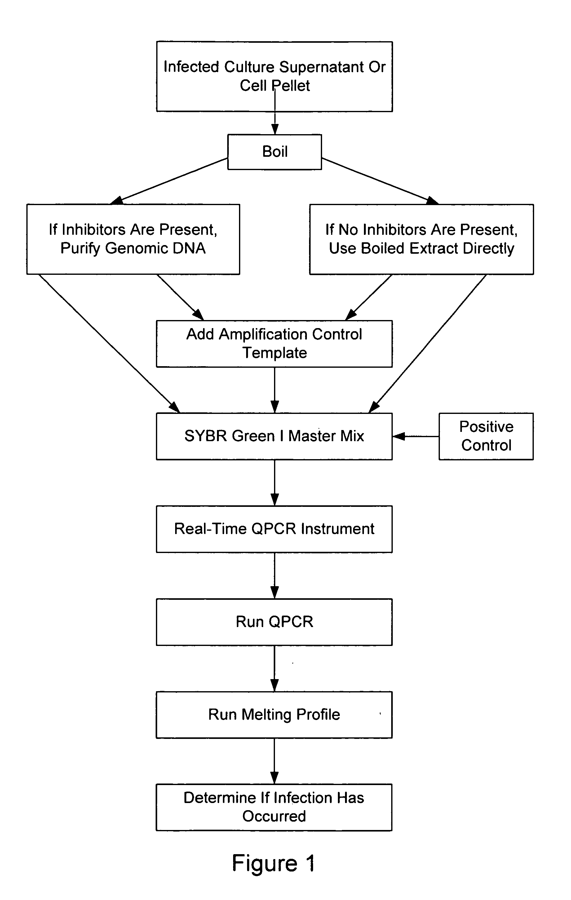
Extraction and Purification of Nucleic Acids from Cell Culture Supernatants

[0138] Cell cultures to be assayed for infection by Mycoplasma / Acholeplasma were grown to near confluence. One hundred microliters (100 μl) of the medium was transferred from the cell culture to a microcentrifuge tube. The tube was tightly closed to prevent opening during subsequent steps, particularly the heating step.

[0139] The microcentrifuge tube was incubated in a water bath at 95-100° C. for 5 minutes, then centrifuged for 30-60 seconds at top speed in a microcentrifuge. The supernatant (approx. 100 μl) was transferred to a new microcentrifuge tube.

[0140] One hundred microliters of the DNA-binding solution provided with the StrataPrep® PCR Purification Kit (Catalog # 400771, Stratagene, La Jolla, Calif.) and 200 μl of 70% (v / v) ethanol were added to the supernatant, and the combination was mixed well. StrataPrep Kit #400711 was used, except the spin cup in the kit were replaced by Stratagene Catalog ...

example 2

Extraction and Purification of Nucleic Acids from Cell Pellets

[0146] As discussed above, cell culture supernatants might contain media components that inhibit PCR amplification (e.g., fetal calf serum, metabolic products, antibiotics). This extraction protocol was used when it was desirable to minimize or eliminate such inhibitory components. It has been found that this protocol provides cell-equivalent standardization and a more sensitive detection limit for cell lines whose growth is inhibited by Mycoplasma. This protocol may be used as an alternative to testing cell culture supernatants that may be inhibitory to PCR.

[0147] Cell cultures to be assayed for infection by Mycoplasma / Acholeplasma were grown to near confluence. The cells were harvested by scraping the cells from the plate without prior addition of trypsin. One ml. of scraped adherent cells (100,000 or more cells) were transferred to a microcentrifuge tube and spun at top speed for 10-15 seconds. The supernatant was ca...

example 3

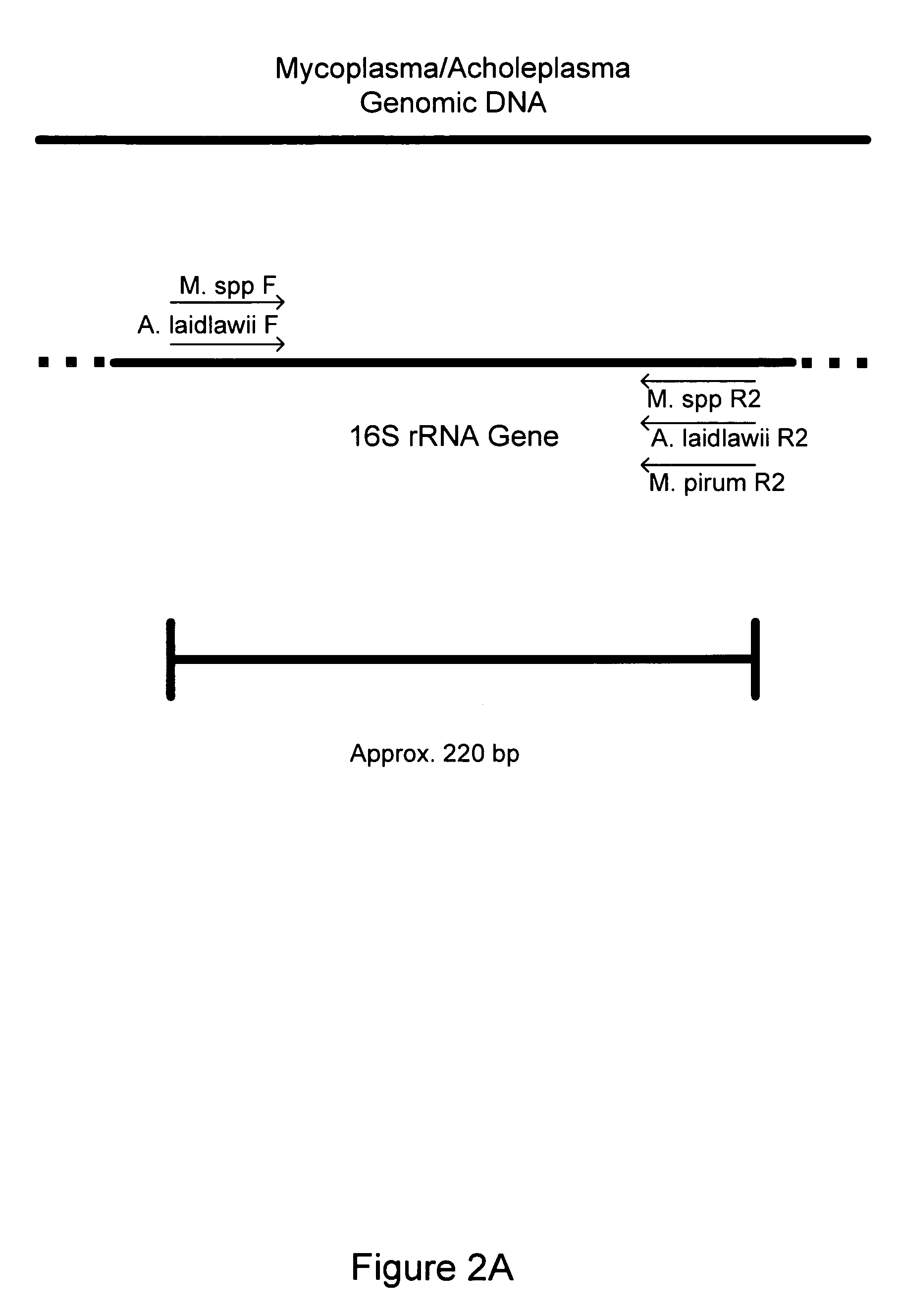
Detection of Eight Different Species of Mycoplasma with One Primer Set

[0157] Eight Mycoplasma or Acholeplasma bacterial strains were obtained as lyophilized preparations from the American Type Culture Collection (ATCC, Manassas, Va.) as follows: Acholeplasma laidlawii (ATCC#23206), Mycoplasma orale (ATCC#29802), M. hominis (ATCC#23114), M. fermentans (ATCC# 19989), M. hyorhinis (ATCC#17981), M. pirum (ATCC#25960), M. salivariuni (ATCC#23064), M. arginini (ATCC#23243). E. coli (ATCC#l 0798) was also obtained for use in determining specificity of the primers. The Mycoplasma / Acholeplasma primer set described in Table 1 was tested for the detection of each of these eight different species of Mycoplasma and E. coli.

[0158] Crude DNA isolation was performed by resuspending lyophilized Mycoplasma bacterial cells in 1 ml of sterile double-deionized (ddI) water, followed by thorough mixing with a aerosol-filtered pipette tip. Resuspension in water ensured hydrolysis of the cells and release...


PUM

PropertyMeasurementUnit
temperatureaaaaaaaaaa
Tmaaaaaaaaaa

Abstract

The invention relates to nucleic acids, methods, compositions, and kits for the PCR-based detection of Mycoplasma and Acholeplasma bacterial species. The nucleic acids, methods, compositions, and kits provide for increased specificity and sensitivity of PCR-based Mycoplasma bacterial assays. Primer sets and PCR-based assays are provided that amplify and detect conserved 16S rRNA gene sequences from multiple Mycoplasma and Acholeplasma species while avoiding amplification and detection of non-target sequences.

Description

FIELD OF THE INVENTION [0001] The invention relates to the detection of bacterial species in biological samples. In particular, the invention relates to detection of nucleic acids specific for Mycoplasma and Acholeplasma species. BACKGROUND OF THE INVENTION [0002] Various species of Mycoplasma and Acholeplasma are involved in human and animal pathologies. Although the first Mycoplasma species was identified in association with bovine pleuropneumonia, it has since been identified as the causative agent of lung disease in humans. Likewise, Acholeplasma species have been implicated in waterfowl, swine, cattle, and human disease. [0003]Mycoplasma contamination of eukaryotic cell cultures is also a common problem, leading to unreliable experimental results and possibly unsafe biological products. Contamination is typically due to presence of Mycoplasma in the original cells used for culture, cross-contamination of laboratory stocks, contamination from compositions added to cell cultures ...

Claims


Application Information

Patent Timeline
Patent Type & Authority Applications(United States)
IPC IPC(8): C07H21/04C12Q1/68
CPCC12Q1/686C12Q1/689C12Q2600/166
Inventor PADMABANDU, GOTHAMISALEHI, FATEMEHPINGERELLI, PETERMUELLER, REINHOLD
Owner STRATAGENE INC US
Why Eureka
Try Eureka