Controlled electroporation and mass transfer across cell membranes in tissue

a cell membrane and cell technology, applied in the field of tissue electroporation, can solve the problems of inability to control the process of electroporation in both individual cells and tissues in real time, low efficiency of transfection by electroporation, and inability to adapt the specific characteristics of individual cells to the current procedure, etc., to achieve the effect of minimizing cellular damage and high efficiency of electrical energy us

Inactive Publication Date: 2005-12-22
RGT UNIV OF CALIFORNIA
View PDF9 Cites 169 Cited by
  • Summary
  • Abstract
  • Description
  • Claims
  • Application Information

AI Technical Summary

Benefits of technology

[0011] The benefits of this process include a high level of control over the onset and degree of electroporation, together with a more detailed knowledge of the occurrence and degree of permeability created in particular cell masses. When applied to cells this process assures that on average, the cells of the tissue are indeed rendered permeable and are indeed transformed by the introduction of chemical species and this can be observed and measured in real time. The process also offers the ability to increase the efficiency of electroporation by avoiding variations in the electrical environment that would destroy some cells while having an insufficient effect on others.
[0019] A feature of the invention is that the magnitude of electrical current during electroporation of cells of tissue becomes dependent on the degree of electroporation of the cells so that current is adjusted within a range predetermined to obtain a desired degree of electroporation minimizing cellular damage.

Problems solved by technology

Unfortunately, the degree of permealization occurring in electroporation varies with the cell type and also varies among cells in a given population.
Furthermore, since the procedure is performed in large populations of cells whose properties vary among the individual cells in the population, the electroporation conditions can only be selected to address the average qualities of the cell population; the procedure as currently practiced cannot be adapted to the specific characteristics of individual cells.
A further problem with known methods of electroporation is that the efficiency of transfection by electroporation can at times be low.
Many of the problems identified above are a consequence of the fact that the process of electroporation in both individual cells and tissues cannot be controlled in real time.
There are no means at present to ascertain in real time when a cell enters a state of electroporation.
As a result, the outcome of an electroporation protocol can only be determined through the eventual consequences of the mass transfer process and its effect on the cell.
This bulk transfer method does not permit precise control or measurement of the mass transfer across the membrane.
Furthermore, the surface of the cell that is exposed to the solution is not well defined.
Cell surface areas vary among cells in a given population, and this leads to significant differences among the cells in the amount of mass transfer.
For these reasons, the amount of mass transfer achieved by bulk transfer processes is not uniform among cells, and the actual amount transferred for any particular cell cannot be determined.
This requires manipulation of the cell with the human hand, a technique that is difficult to perform, labor-intensive, and not readily reproducible.
Electroporation with microelectrodes suffers these problems as well as the lack of any means to detect the onset of electroporation in an individual cell.

Method used


Image

  • Controlled electroporation and mass transfer across cell membranes in tissue
  • Controlled electroporation and mass transfer across cell membranes in tissue
  • Controlled electroporation and mass transfer across cell membranes in tissue

Examples


example

Numerical Method

[0086] A simplified two-dimensional isotropic cross-section of a liver 7 cm in diameter is used for the simulation. The surface of the analyzed tissue is assumed to be electrically isolated with a zero flux boundary condition to simulate a situation in which the liver is temporarily placed in an insulating, semi-rigid electrode array fixture during a surgical procedure. To collect data for the reconstruction algorithm, 32 electrodes are equally spaced around the periphery of the virtual phantom, i.e. electroporation conductivity map.

Experimental Results used in Front-Tracking Reconstruction Algorithm

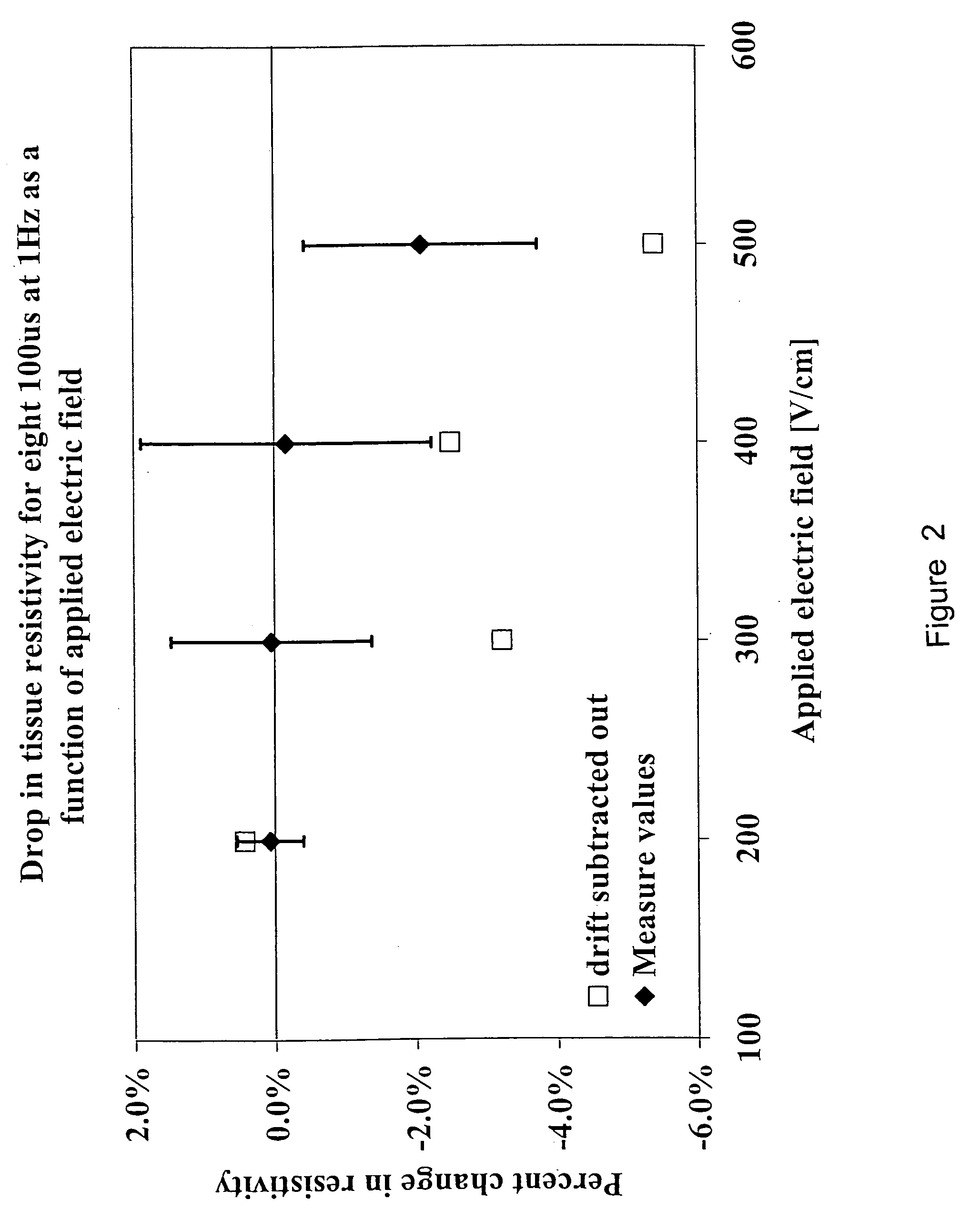
[0087] For computational reasons and for model demonstration, we used the data from the 10 ms pulses in this study. Specifically, with these experiments, eight 10 ms pulses at a frequency of 1 Hz were delivered for electric fields of 0, 200, 300, 400 and 500V cm″1. The threshold gradient required to induce electroporation was taken from the experimental data and was sh...

example 2

Electroporation Model

[0088] The electroporation simulation evaluates the potential and gradient distribution in the tissue due to an electrical pulse using the finite element method. To account for the change in electrical properties of the tissue during electroporation, the algorithm iteratively solves the Poisson equation with a zero source term:

Δ·(σΔφ)=0

[0089] where σ is the electrical conductivity and φ is the electrical potential. The conductivity of the tissue is assumed to be initially at 0.13S m−1, which agrees with results for dog liver in situ at IkHz and measured from one sample. At locations where the voltage gradient surpasses a threshold gradient, the conductivity increases as prescribed by experimental results in FIG. 10. The gradients are recalculated with the updated conductivity values until the solution converges. For convergence, it was assumed that once the conductivity of an element increases, it will not revert to a lower value, at least within the duration ...

example 3

Ex Vivo Reconstruction Examples

[0090] In addition to the baseline example, three electroporation electrode geometrical configurations were investigated, which are modeled after in vivo electroporation experiments conducted on rabbit liver and murine skeletal muscle.

[0091] The first configuration which is show in FIG. 11 had two electrodes separated with an inner distance of 8 mm, centered in the liver, with one electrode at 800V and the other grounded. The second configuration was similar to the first, except that the applied voltage was 600V. The third configuration had two rows of four electrodes centered in the liver. The center-to-center distance between the two rows was 4 mm, and the center-to-center spacing between the electrodes in each row was 2 mm. The voltage of the top row was set to 1200V and the bottom row to ground. The diameters of the electrodes for the two examples were I.imm, 0.3 mm, and 0.5 mm, respectively.


PUM

PropertyMeasurementUnit
porosityaaaaaaaaaa
thicknessaaaaaaaaaa
frequencyaaaaaaaaaa

Abstract

Electroporation is performed in a controlled manner in individual and multiple biological cells present in biological tissue by monitoring the electrical impedance, defined herein as the ratio of current to voltage in the electroporation cell. The impedance detects the onset of electroporation in the biological cell(s), and this information is used to control the intensity and duration of the voltage to assure that electroporation has occurred without destroying the cell(s). This is applicable to electroporation in general.

Description

CROSS-REFERENCE [0001] This application is a continuation-in-part of application Ser. No. 10 / 079,940, filed Feb. 19, 2002 which is a continuation-in-part of application Ser. No. 09 / 618,951, filed Jul. 19, 2000 which is a continuation-in-part of application Ser. No. 09 / 358,510, filed Jul. 21, 1999, now U.S. Pat. No. 6,300,108 issued Oct. 9, 2001, all of which are incorporated herein by reference and to which are claimed priority under 35 USC §120.FIELD OF THE INVENTION [0002] This invention resides in the fields of electroporation and mass transfer across cell membranes and more specifically to electroporation of cells in tissue. BACKGROUND OF THE INVENTION [0003] Electroporation is a technique that is used for introducing chemical species into biological cells, and is performed by exposing the cells to an electric potential that traverses the cell membrane. While its mechanism is not fully understood, electroporation is believed to involve the breakdown of the cell membrane lipid bi...

Claims


Application Information

Patent Timeline
Patent Type & Authority Applications(United States)
IPC IPC(8): C12N15/87
CPCA61N1/0412A61N1/0476C12N15/87A61N1/3754C12M35/02A61N1/327
Inventor RUBINSKY, BORISHUANG, YONG
Owner RGT UNIV OF CALIFORNIA
Why Eureka
Try Eureka