
Hand-held imaging probe for treatment of states of low blood perfusion

a handheld imaging and state-of-low blood perfusion technology, applied in the field of non-invasive handheld imaging instruments, can solve the problems of low success rate of thrombolytic drug treatment, marked decline in in-hospital survival, burning of overlying skin and soft tissue, etc., and achieves advantageous sized and shaped, high energy acoustic energy, and substantially rigidity.

Inactive Publication Date: 2006-02-02
AHOF BIOPHYSICAL SYST
View PDF92 Cites 21 Cited by
  • Summary
  • Abstract
  • Description
  • Claims
  • Application Information

AI Technical Summary

Benefits of technology

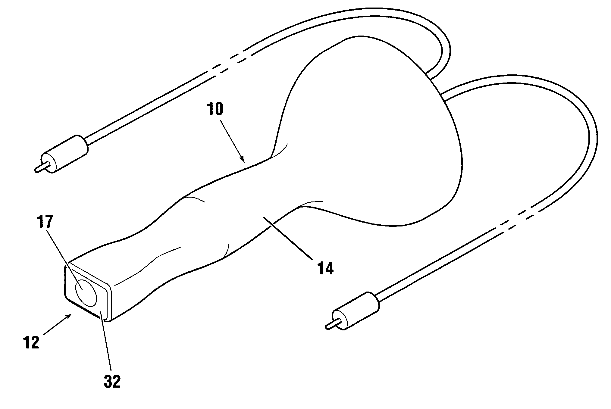
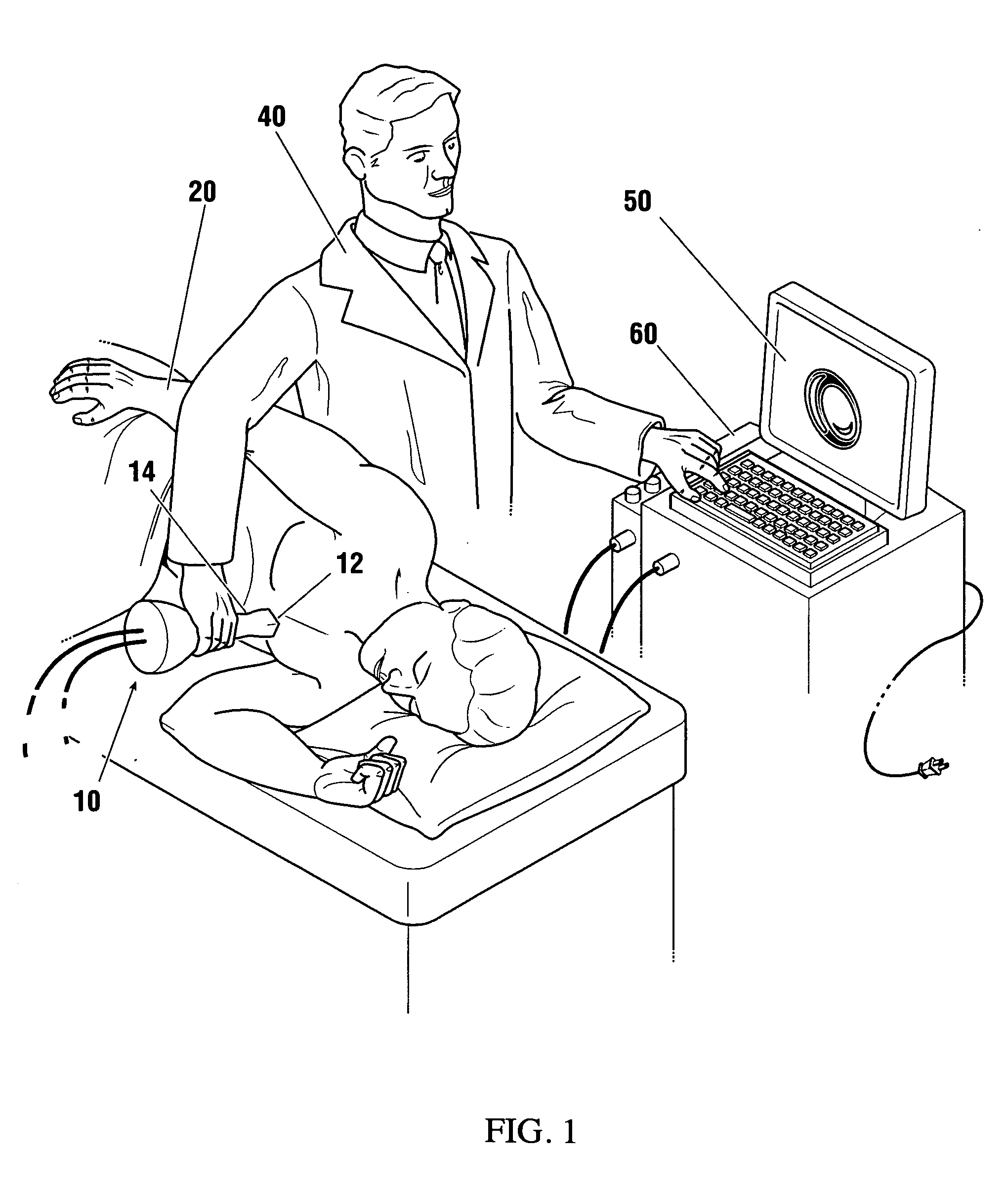
[0016] The present invention provides a non-invasive hand-held treatment imaging probe, which enables the therapeutic delivery of high intensity, high sonic to low ultrasonic frequency acoustic energy to a targeted vascular region, while under ultrasonic imaging guidance via an established acoustic energy penetration window. The treatment imaging probe advantageously comprises a combination of an ultrasonic imaging transducer, and, preferably, a phased array imaging transducer, operatively attached to a therapeutic actuator, the combination configured and sized for hand-held use. In the preferred embodiment, an engagement face of the ultrasonic imaging transducer is operatively disposed about an application end of the therapeutic actuator (operable to emit high energy acoustic energy in about the 1-500 kHz range, and preferably 1-150 kHz range, and most preferably 15-30 kHz range), such that the engagement face and application end of both the ultrasonic imaging transducer and the therapeutic actuator, respectively, share a common application surface on the treatment imaging probe. The application surface of the treatment imaging probe is substantially rigid, and advantageously sized and shaped to enable efficient seating within an intercostal space of a patient, thereby enabling the targeted delivery of high intensity, therapeutic acoustic energy via ultrasonic imaging guidance to the chest wall in thoracic cavity applications. The treatment imaging probe is advantageously designed such that the application surface will not appreciably overheat, and cause burning of the overlying skin or soft tissue of a patient receiving therapy.
[0020] As stated, the preferred therapy is combined with systemically administered clot disruptive and vasodilatory drug therapy, and / or cavitating spheres or microbubbles to accentuate the internal oscillative effect, whereby the therapeutic acoustic effects assist clot disruption, vasodilation, and improved drug interaction and effectiveness to the targeted vascular region. It should be understood that the therapeutic high sonic to low ultrasonic acoustic energy may be imparted continuously, or may be provided in pulses, preferably with a selectable duty factor, in accordance with the desired therapy.
[0024] It is a further purpose of the present invention to provide a treatment imaging probe of the aforementioned type, comprising an ultrasonic imaging transducer operatively attached to a therapeutic actuator operable to emit acoustic energy in about the 1-500 kHz range, and preferably in the 1-150 kHz range, and most preferably in about the 15-30 kHz range, such that the application end of the therapeutic actuator and the engagement face of the ultrasonic imaging transducer share a substantially common application surface, thereby enabling a simultaneous application of high intensity, high sonic to low ultrasonic therapeutic acoustic energy with directed ultrasonic imaging, through an established acoustic penetration window.
[0026] It is a further purpose of the present invention to provide a treatment imaging probe of the aforementioned type, wherein the application surface of the instrument is substantially rigid and sized to enable efficient seating within a rib space of a patient, such as to enable optimized imaging techniques and maximized penetration of acoustic energy in thoracic cavity applications.
[0028] It is a further purpose of the present invention to provide a treatment imaging probe of the aforementioned type, which has means to prevent overheating of the application surface to avoid potential burning of the overlying soft tissue and skin of a patient during therapy.

Problems solved by technology

Thrombolytic drug treatment does not, however, have a high success rate, with adequate reperfusion occurring only between 50-63% of the time within ninety minutes of administration of the thrombolytic drug.
Furthermore, success with drug based reperfusion treatment and in-hospital survival declines markedly when the patient becomes hemodynamically unstable or enters cardiogenic shock, which is the leading cause of in-hospital deaths from AMI in North America.
These higher intensity requirements have tended to cause burning of overlying skin and soft tissue, and have thereby been problematic in practical use.
It is well known however that overlying body tissue often blocks penetration of the acoustic waves as the waves are absorbed as heat or reflected and dissipated throughout the overlying body tissue, and thereby often never reach their intended vascular target, which is more deeply situated, especially in cardiac applications where the interference of dense overlying intercostal tissue and overlying lung does not transmit acoustic energy effectively.
Furthermore the actuator disclosed by Siegel is sub-optimal as it does not include a mechanism to prevent burning of the patient's skin, which is a common concern in high intensity, low ultrasonic frequency skin surface delivery.
Piezoelectric crystals and ultrasonic transmission systems designed for ultrasonic imaging operate in the higher frequency megahertz range, thus the therapeutic pulses are severely limited in penetration power and therapeutic effectiveness.
None of the previously known instruments have a suitable application surface specifically sized and structured to enable efficient seating within a rib space of a patient, which can be of special importance to achieve optimal penetration in thoracic cavity, or cardiological applications, and none of the previously disclosed systems are equipped with the appropriate combination of high intensity emission capabilities, and means for limiting the heating of the contact surface, such as is required for external skin surface applications where deep penetration is generally required.
Lithotripters require precise focussing of a target which is not possible in coronary thrombosis applications, as the coronary arteries cannot be imaged by non-invasive ultrasonic techniques, and the culprit blood clot thereby comprises a hidden, moving target.
Furthermore, lithotripters are not designed to enable continuous wave, or high duty factor pulsed wave, acoustic therapy via a purposively divergent beam, hence the targeted area and prospective effectiveness of these treatment systems are substantially limited.

Method used


Image

  • Hand-held imaging probe for treatment of states of low blood perfusion
  • Hand-held imaging probe for treatment of states of low blood perfusion
  • Hand-held imaging probe for treatment of states of low blood perfusion

Examples


Embodiment Construction

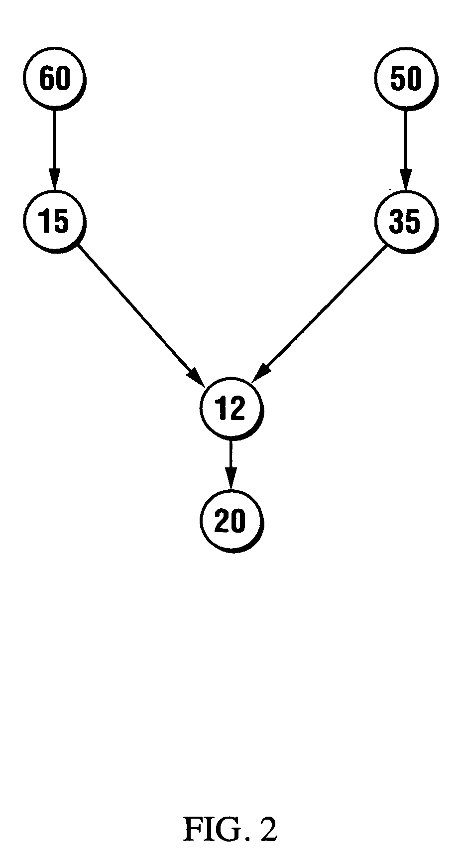
[0036] A non-invasive hand-held treatment imaging probe 10 for treating emergency blood flow disturbances and states of low blood perfusion to body regions by imparting therapeutic high sonic to low ultrasonic frequency acoustic energy is described. Treatment imaging probe 10 comprises an ultrasonic imaging transducer 35 operatively attached to a therapeutic actuator 15. The therapeutic actuator 15 is operable in about the 1-500 kHz, and preferably 1-150 kHz, and most preferably 15-30 kHz, frequency range. As stated previously, therapeutic acoustic energy in the high sonic to low ultrasonic frequency ranges is known for its deep penetration characteristics, and superior clot disruptive and thrombolytic enhancement capabilities. Treatment imaging probe 10 has a substantially rigid application surface 12 which is generally sized and shaped to enable seating within a rib space of a patient 20. Application surface 12 advantageously includes an engagement face 32 of ultrasonic imaging tr...


PUM


Abstract

A non-invasive hand-held treatment imaging probe for treatment of acute blood flow disturbances and states of low blood perfusion. The treatment imaging probe is operable to emit high intensity therapeutic mechanical acoustic waves while under ultrasonic imaging guidance. The probe has a substantially rigid application surface, sized to enable seating within a rib space of a patient (i.e. for cardiac applications), comprising the combination of an engagement face of an ultrasonic imaging transducer and an application end of a high powered therapeutic actuator operable in about the 1-500 kHz (and preferably 1-150 kHz) range. Treatment imaging probe can be used as an adjunct to thrombolytic therapy in the treatment of acute thrombotic vascular obstructions, such as in acute myocardial infarction, or alternatively to enhance localized delivery of angiogenic agents or other useful medications to targeted vascular regions.

Description

CROSS-REFERENCE TO RELATED APPLICATIONS [0001] This application is a continuation-in-part of co-pending U.S. patent application Ser. No. 10 / 902,122, filed Jul. 30, 2004, the contents of which are expressly incorporated herein by reference.FIELD OF THE INVENTION [0002] This invention relates to non-invasive hand-held imaging instruments adapted to impart therapeutic acoustic energy to a human body, for emergency treatment of acute blood flow disturbances. BACKGROUND OF THE INVENTION [0003] Acute thrombosis, ischemia and coronary artery disease are all common concerns. Acute myocardial infarction (AMI) subsequent to coronary thromboses, in particular, is one of the leading causes of death in North America and Europe. Current first-line treatment of thromboses in the acute phase when the patient reaches professional care is typically by intravenous administration of thrombolytics, or a combination of drugs such as heparin, aspirin and / or GP 2b 3a platelet inhibitors to dissolve the blo...

Claims


Application Information

Patent Timeline
IPC IPC(8): A61B8/12
CPCA61B8/08A61B8/0883A61N7/00A61B8/4455A61B8/483A61B8/0891
Inventor HOFFMANN, ANDREW KENNETH
Owner AHOF BIOPHYSICAL SYST
Learn More