
Devices and methods of screening for neoplastic and inflammatory disease

a technology of inflammatory disease and devices, applied in the field of devices and methods of screening for inflammatory disease, can solve the problems of inability to detect, inability to detect, and long detection time, and achieve the effect of reducing inflammation or control and being convenient to us

Inactive Publication Date: 2006-02-16
HELLER ADAM
View PDF68 Cites 44 Cited by
  • Summary
  • Abstract
  • Description
  • Claims
  • Application Information

AI Technical Summary

Benefits of technology

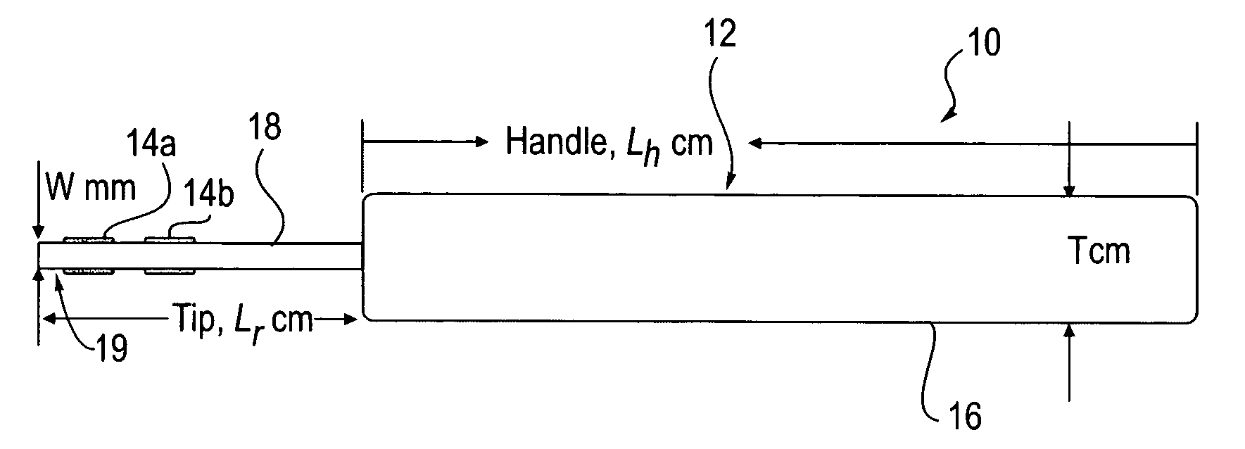
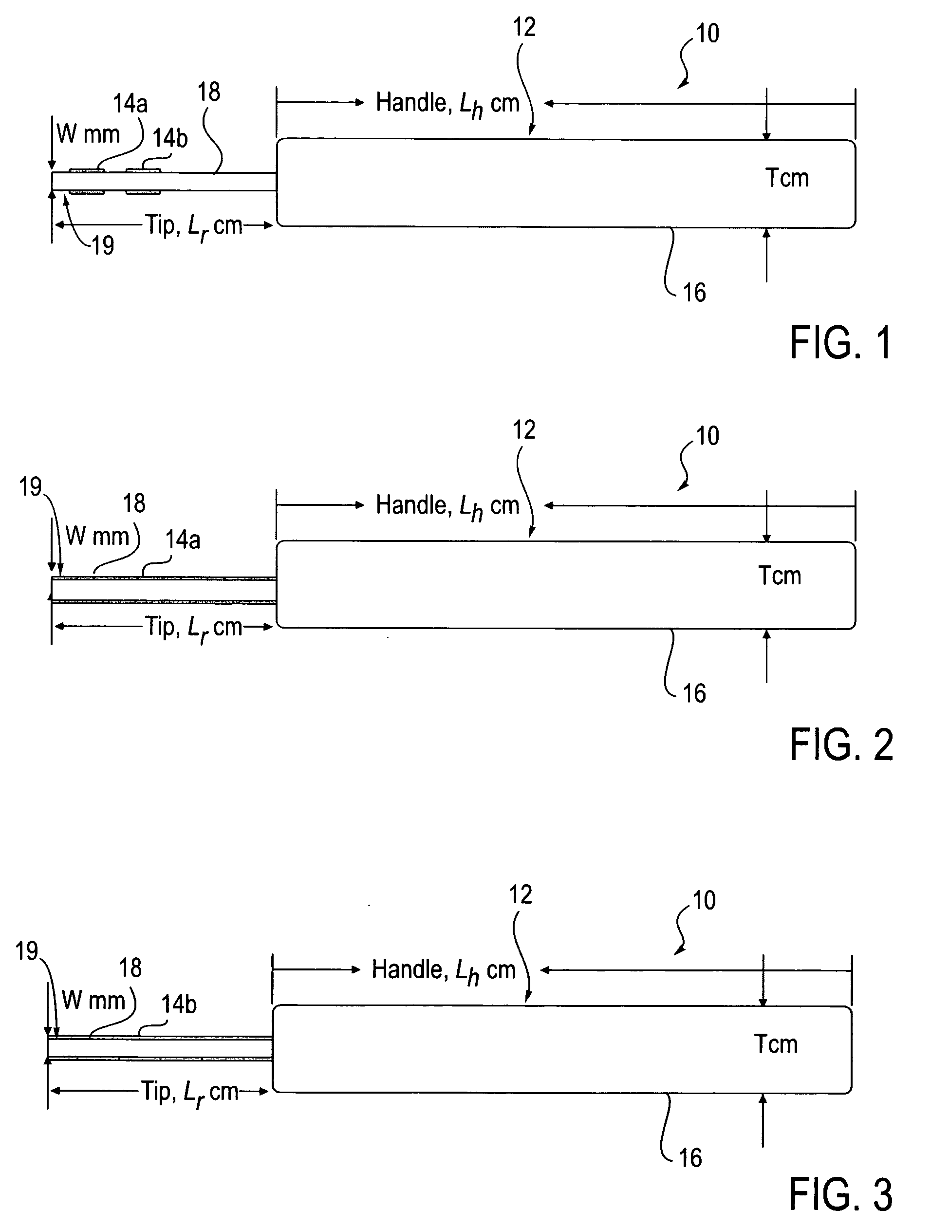
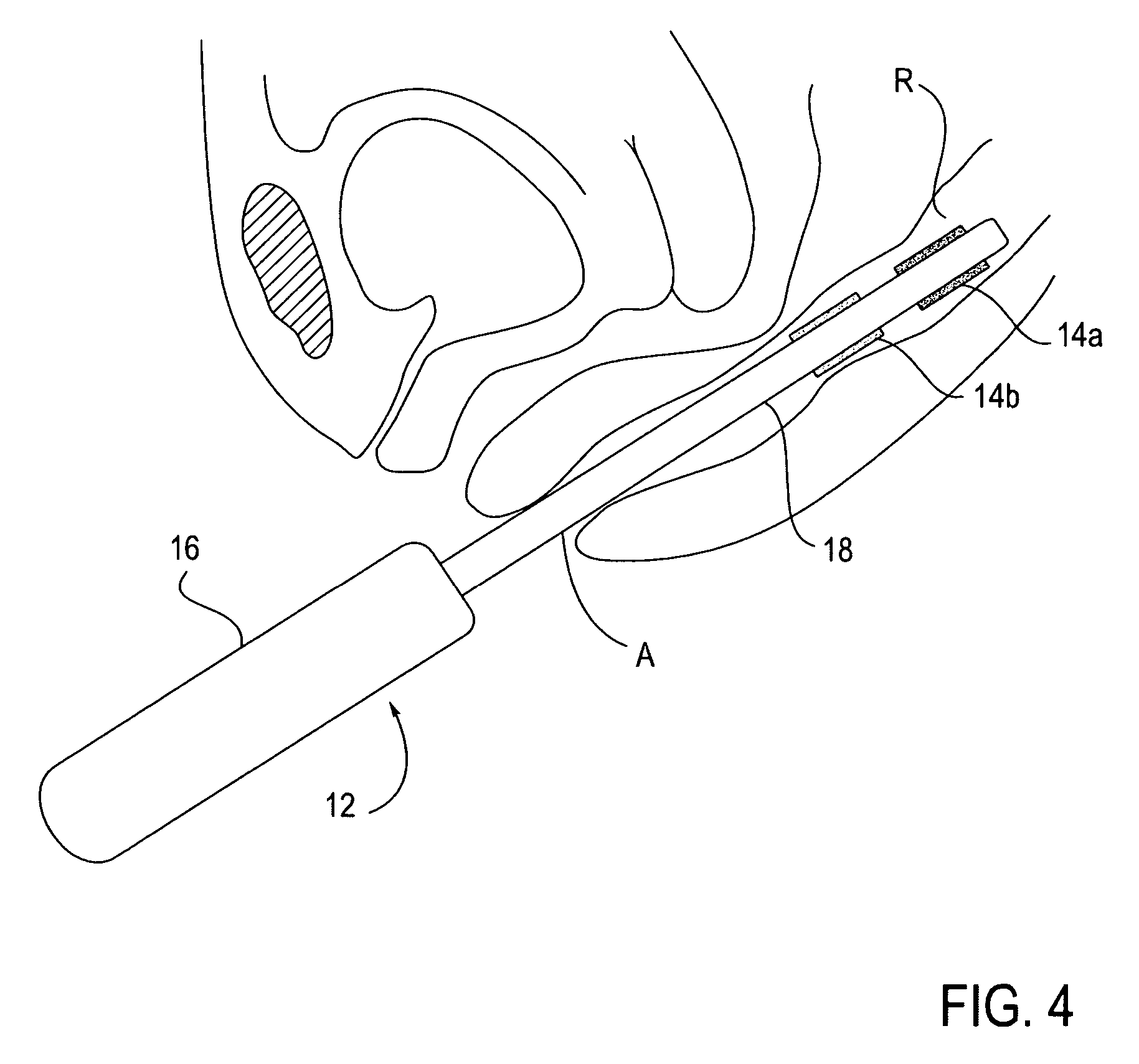
[0067] Methods and devices are provided for evaluating the presence of disease in a patient. In particular, methods and devices provided for screening patients for neoplastic and inflammatory disease. Such diseases are often indicated by the elevated level of a chemical compound associated with disease, particularly nitric oxide (NO) and / or nitrogen dioxide (NO2). Through measuring and / or estimating the chemical compound-concentration, the methods and tools provided distinguish between patients who require further testing and / or treatment and those who do not. The methods and tools also provide information about the effectiveness of treatment, such as treatment to reduce inflammation or control of the growth of malignant tumors. These methods and devices are relatively inexpensive, easy to use, and provide many advantages which are described herein.

Problems solved by technology

Although NO has an unpaired electron and is a free radical, its half life, in absence of a catalyst of its oxidation by molecular oxygen, is very long.
Although NO analyzing diagnostic methods have been available, some for many years, the known methods required expensive instrumentation, required trained professionals to operate the instruments, involved systems that are much too heavy and too large to be carried by the patient or the physician, and were, as is evident from the literature cited, far from simple to use.
This delayed receipt of the information and the method provided little or no information about the tissue, organ or specific volume element of the body in which the NO-concentration was elevated.

Method used


Image

  • Devices and methods of screening for neoplastic and inflammatory disease
  • Devices and methods of screening for neoplastic and inflammatory disease
  • Devices and methods of screening for neoplastic and inflammatory disease

Examples


example 1

Probe Materials in Which Blue PTIO was Bleached by NO

[0198] 2-phenyl-4,4,5,5-tetramethylimidazoline-1-oxyl 3-oxide (PTIO) was purchased fro Sigma-Aldrich Corp., St. Louis, Mo. FDA (a) White silicone rubber probe, 0.093 inches in diameter part # SC6020204 was purchased from Ipotec Inc., Exeter, N.H. The part of the probe to be dyed was pre-soaked for 10 min in tetrahydrofuran (THF). About 6 mg of the PTIO were dissolved in about 10 mL of THF. The pre-soaked probe was then immersed in the PTIO solution for 5 min and allowed to dry for 24 h. Upon soaking, the silicone rubber turned blue. Its blue NO-reactive material was not removed by wiping, even when the wiping tissue was wetted with THF. Nitric oxide was generated in a vial by mixing equal volumes of aqueous solutions of about 1 M FeSO4 and about 0.5 M NaNO2. An about 1″ long segment of the dyed part of the probe was exposed for about 20 s to the NO-containing gas. The exposed segment was bleached, loosing its blue color. (b) As i...

example 2

[0199] The paper tape of Example 1 (e) was adhered to an inflamed cut in the skin of a volunteer for about 10 hours. The inflamed region of the cut was precisely mapped and was clearly visible as a colorless domain in the blue tape.

example 3

[0200] The paper tape of Example 1 (e) was adhered to the front end part of an endoscope probe and applied in colonoscopy. The paper turned colorless in the typically 1-3 minute long procedure in patients with inflammatory bowel disease revealed by parallel colonoscopy or endoscopy.


PUM


Abstract

Methods and devices are provided for evaluating the presence of disease in a patient. In particular, methods and devices are provided for screening patients for neoplastic and / or inflammatory disease. Such diseases are often indicated by the elevated level of a chemical compound associated with disease, such as nitric oxide (NO) and / or nitrogen dioxide (NO2). Through measuring and / or estimating the chemical compound-concentration, such as by change in fluorescence, absorbance or reflectance, the methods and tools provided distinguish between patients who require further testing and / or treatment and those who do not. The methods and tools also provide information about the effectiveness of treatment, such as treatment to reduce inflammation or control of the growth of malignant tumors. These methods and devices are relatively inexpensive, easy to use, and provide other advantages.

Description

CROSS-REFERENCES TO RELATED APPLICATIONS [0001] This application claims the benefit and priority of U.S. Provisional Patent Application No. 60 / 599,752 (Attorney Docket 021821-000500US), filed Aug. 6, 2004, and U.S. Provisional Patent Application No. 60 / 683,518 (Attorney Docket 021821-000520US), filed May 20, 2005, the full disclosures of which are hereby incorporated by reference for all purposes.STATEMENT AS TO RIGHTS TO INVENTIONS MADE UNDER FEDERALLY SPONSORED RESEARCH OR DEVELOPMENT [0002] NOT APPLICABLE REFERENCE TO A “SEQUENCE LISTING,” A TABLE, OR A COMPUTER PROGRAM LISTING APPENDIX SUBMITTED ON A COMPACT DISK [0003] NOT APPLICABLE BACKGROUND OF THE INVENTION [0004] There is a need for clinically useful methods and tools for diagnosing pathological elevation in the concentration of nitric oxide (NO) in parts of the human body. NO is a recognized endothelium derived relaxing factor, a cardiovascular signaling molecule and a neurotransmitter. It is found in most, possibly in al...

Claims


Application Information

Patent Timeline
Patent Type & Authority Applications(United States)
IPC IPC(8): A61B5/00
CPCA61B5/145A61B5/4547A61B5/4283A61B5/411
Inventor HELLER, ADAMLIEBERMANN, THOMAS
Owner HELLER ADAM
Learn More