
Biomolecule analysis using Raman surface scanning

Inactive Publication Date: 2006-03-02
INTEL CORP
View PDF23 Cites 52 Cited by
  • Summary
  • Abstract
  • Description
  • Claims
  • Application Information

AI Technical Summary

Problems solved by technology

When this principle is applied to increase efficiencies of biochemical or clinical analyses, the principal challenge is to develop a probe identification system that has distinguishable components for an individual probe in a large probe set.
In addition quantum dots that emit at short wavelength need UV excitation when used in highly multiplexed detection and such UV excitation creates a problem of background signal.
Either method requires complicated instrumentation to fabricate the arrays, may accommodate a relatively low probe density, and does not afford the user the option of changing the contents of the chip.
Similarly, conventional cell imaging techniques utilizing fluorescent dyes suffer from toxicity of fluorescent chemicals (which often kill the cells), limited lifetime of dyes (which bleach over time), and limited number of colors that may be used together due to the broad dye emission spectra of fluorescent dye molecules.
However, quantum dots raise environmental and safety issues due to use of cadmium core material.
Traditional methods for protein profiling, such as two-dimensional gel and MDLC-MS (Multi-dimensional Liquid Chromatographs-Mass Spectroscopy), may not detect low-abundance proteins, such as those present at a concentration less than 1 ng / mL and requires isolation of the samples from their source, leading to loss of valuable information regarding features of the sample.
None of the foregoing techniques is well suited for providing quantitative measurements, however.
Consequently SERS has not gained widespread use.
In addition, many biomolecules such as proteins and nucleic acids do not have unique Raman signatures because these types of molecules are generally composed of a limited number of common monomers.

Method used


Image

  • Biomolecule analysis using Raman surface scanning
  • Biomolecule analysis using Raman surface scanning
  • Biomolecule analysis using Raman surface scanning

Examples


example 1

[0146] Antibody-COIN conjugation: To conjugate COIN particles with antibodies, a direct adsorption method was used. A 500 μL solution containing 2 ng of a biotinylated anti-human IL-2 (anti-IL-2), or IL-8 antibody (anti-IL-8), in 1 mM Na3Citrate (pH 9) was mixed with 500 μL of a COIN solution (using 8-aza-adenine or N-benzoyl-adenine as the Raman label); the resulting solution was incubated at room temperature for 1 hour, followed by adding 100 μL of PEG-400 (polyethylene glycol 400). The solution was incubated at room temperature for another 30 min before a 200 μL of 1% Tween-20 was added. The resulting solution was centrifuged at 2000×g for 10 min. After removing the supernatant, the pellet was resuspended in 1 mL solution (BSAT) containing 0.5% BSA, 0.1% Tween-20 and 1 mM Na3Citrate. The solution was again centrifuged at 1000×g for 10 min to remove the supernatant. The BSAT washing procedure was repeated for a total of 3 times. The final pellet was resuspended in 700 μL of Diluti...


PUM


Abstract

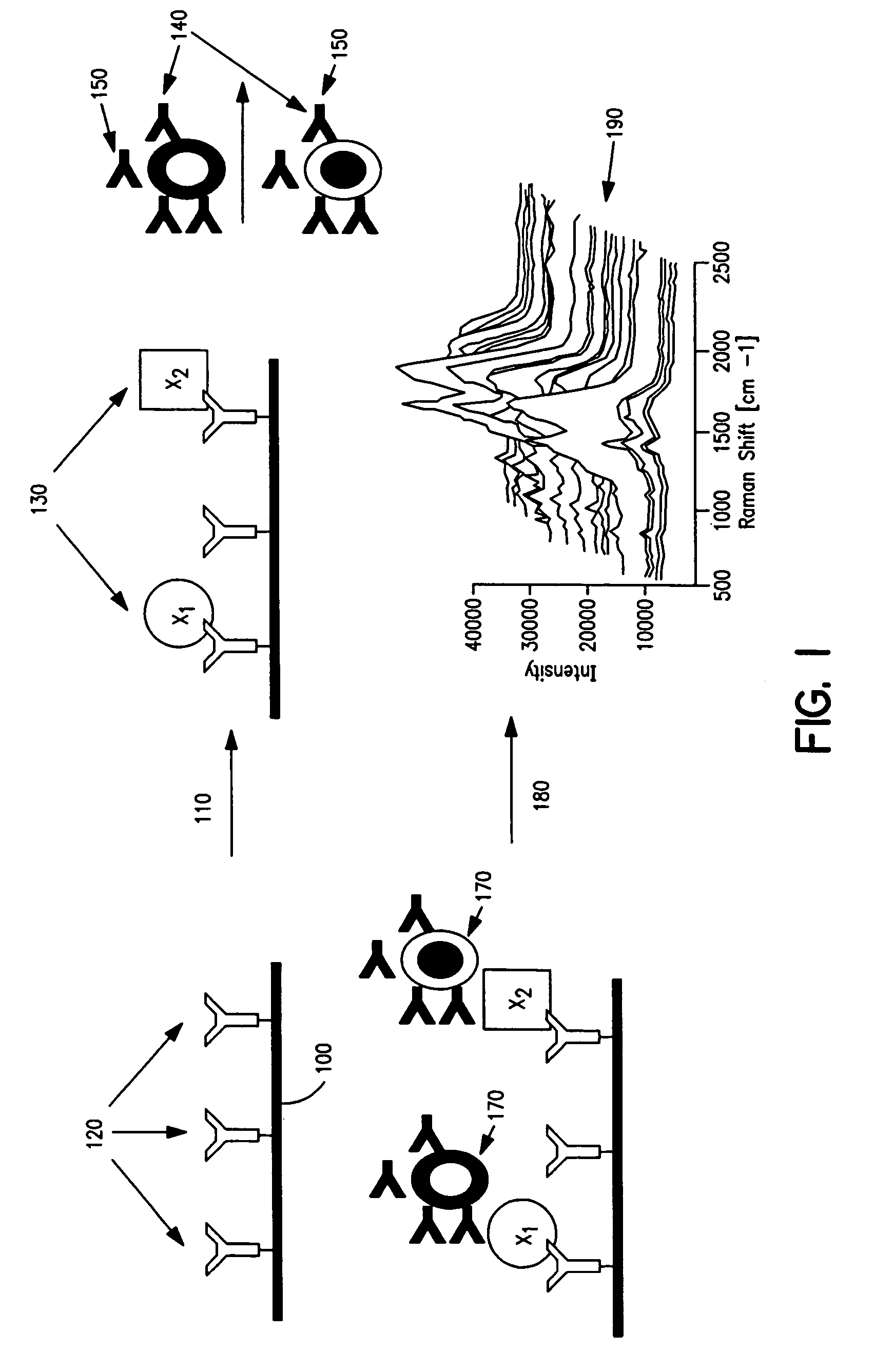
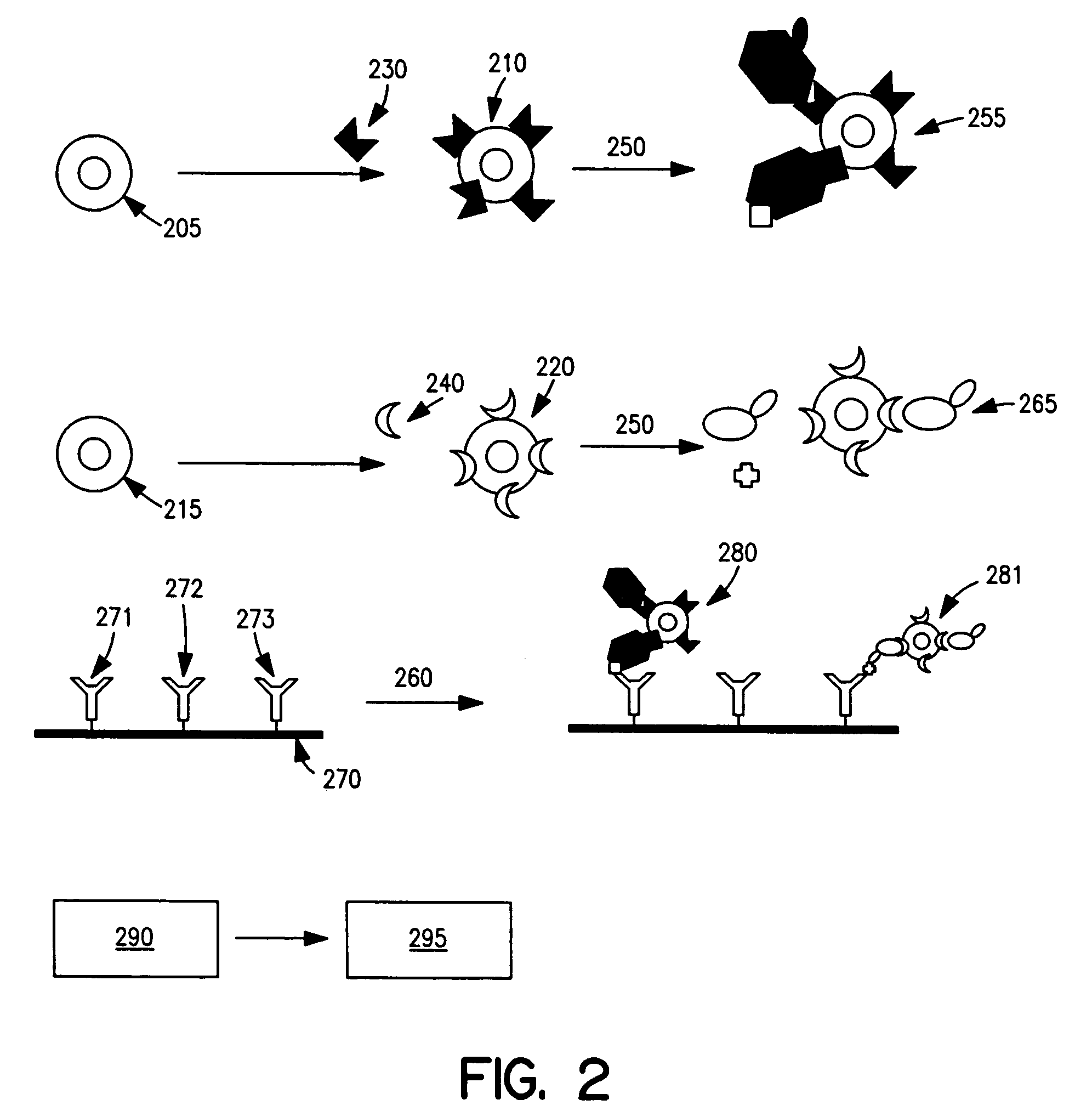
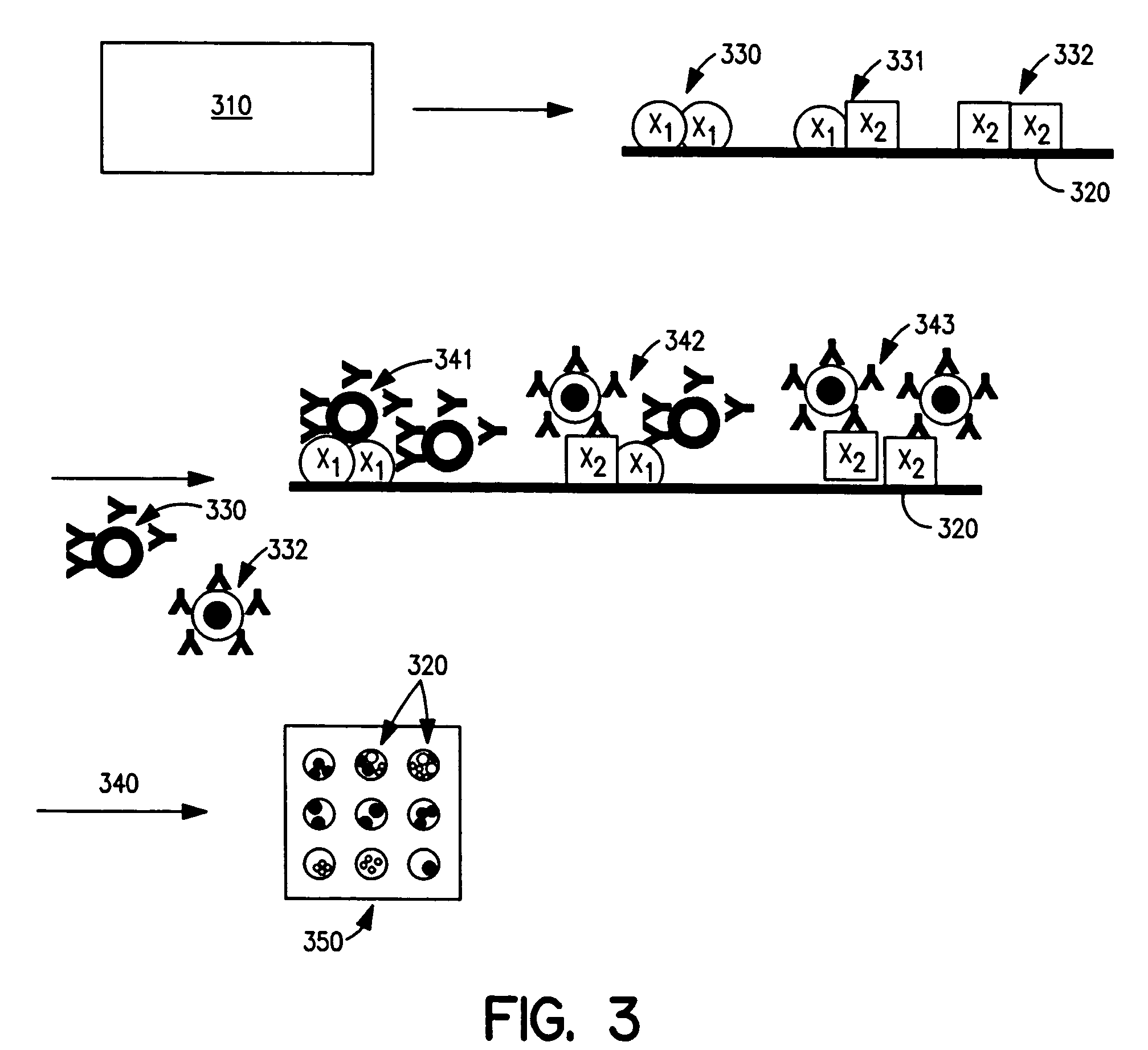
Methods and apparatus are provided herein for assaying biological samples using probes labeled with composite organic-inorganic nanoparticles (COINs) and microspheres with COINs embedded within a polymer matrix to which the probe moiety is attached. COINs are Raman-active nanoparticles made up of aggregated primary metal crystal particles with Raman-active organic compounds adsorbed on the surface in the junctions of aggregated primary metal crystal particles or embedded in the crystal lattice of the primary metal particles. Since COINs intrinsically produce SERS signals upon laser irradiation, COIN-labeled probes are particularly suitable in a variety of methods for assaying biological molecules, most of which are not inherently Raman-active. The invention provides variations of the sandwich immunoassay employing both specific and degenerate binding, methods for reverse phase assay of tissue samples and cell microstructures, in solution displacement and competition assays, and the like. Kits and chips useful for practicing the invention assays are also provided.

Description

BACKGROUND OF THE INVENTION [0001] 1. Field of the Invention [0002] The invention relates generally to use of nanoparticles for biomolecule analysis, and more specifically to the use of such nanoparticles in biomolecule analysis by surface-enhanced Raman spectroscopy. [0003] 2. Background Information [0004] Multiplex reactions are parallel processes that exist naturally in the physical and biological worlds. When this principle is applied to increase efficiencies of biochemical or clinical analyses, the principal challenge is to develop a probe identification system that has distinguishable components for an individual probe in a large probe set. High-density DNA chips and microarrays are probe identification systems in which physical positions on a solid surface are used to identify nucleic acid or protein probes. The method of using striped metal bars as nanocodes for probe identification in multiplex assays is based on images of the metal physical structures. Quantum dots are par...

Claims


Application Information

Patent Timeline
IPC IPC(8): G01N33/543
CPCG01N33/54366G01N33/58G01N33/54373
Inventor SUN, LEI B.SU, XINGKOO, TAE-WOONGCHAN, SELENA
Owner INTEL CORP
Learn More