Hydrocolloid coating of a single cell or embryo

Inactive Publication Date: 2006-03-23
YISSUM RES DEV CO OF THE HEBREW UNIV OF JERUSALEM LTD
View PDF5 Cites 19 Cited by
  • Summary
  • Abstract
  • Description
  • Claims
  • Application Information

AI Technical Summary

Benefits of technology

[0019] The present invention discloses for the first time the unexpected findings that the hydrocolloid coating of the cell or embryo: (a) extended survival rates, (b) protected the cell or embryo from pathogen contamination, (c) protected the cell or embryo from hazardous materials produced or introduced into the

Problems solved by technology

These preparations regularly suffer from low mechanical strength and possible heat damage.
The limitations of such systems are low mechanical strength and breakdown in the presence of chelating agents.
However, the above-identified patents teach multi-cellular entrapping and not a single cell coating.
However, this patent teaches multi-cellular coating, and coating a single viable cell (e.g. an egg) or embryo is neither taught nor suggested.
Aquaculture raised fish are vulnerable to infections.
Losses to stock from these infections reduce productivity and increase consumer costs, greater than $100 million dollars each year.
Fish embryos are susceptible to a variety of bacterial infections that can have a devastating effect on the stock of a fish farm.
Embryos and hatchlings cannot

Method used


Image

  • Hydrocolloid coating of a single cell or embryo
  • Hydrocolloid coating of a single cell or embryo
  • Hydrocolloid coating of a single cell or embryo

Examples


example 1

The Adhesion Properties of X. laevis Eggs and Embryos to Different Substrates

[0081] The adhesion properties of Xenopus laevis eggs and embryos to various surfaces (substrates) were determined in different experimental set-ups. They were divided into experiments conducted on nonfertilized and fertilized eggs (embryos). The nonfertilized eggs were examined immediately after ovulation of the eggs, after swelling of the jelly coat, and after different periods of time has elapsed from the moment of adhesion. For the fertilized eggs, adhesion was examined after swelling of the jelly coat and 1 h after fertilization.

[0082] The roughness of the five hydrocolloid-gel systems (agarose, agar, alginate, κ-carrageenan, and gelatin) could be estimated by atomic force microscopy, gloss measurement, or by sensory evaluation as highly smooth surfaces. It is important to note that these hydrocolloids differ in their compositions, structure, and overall properties (Nussinovitch, 1997, Hydrocolloid A...

example 2

Hydrocolloid Coating of X. laevis Eggs Embryos

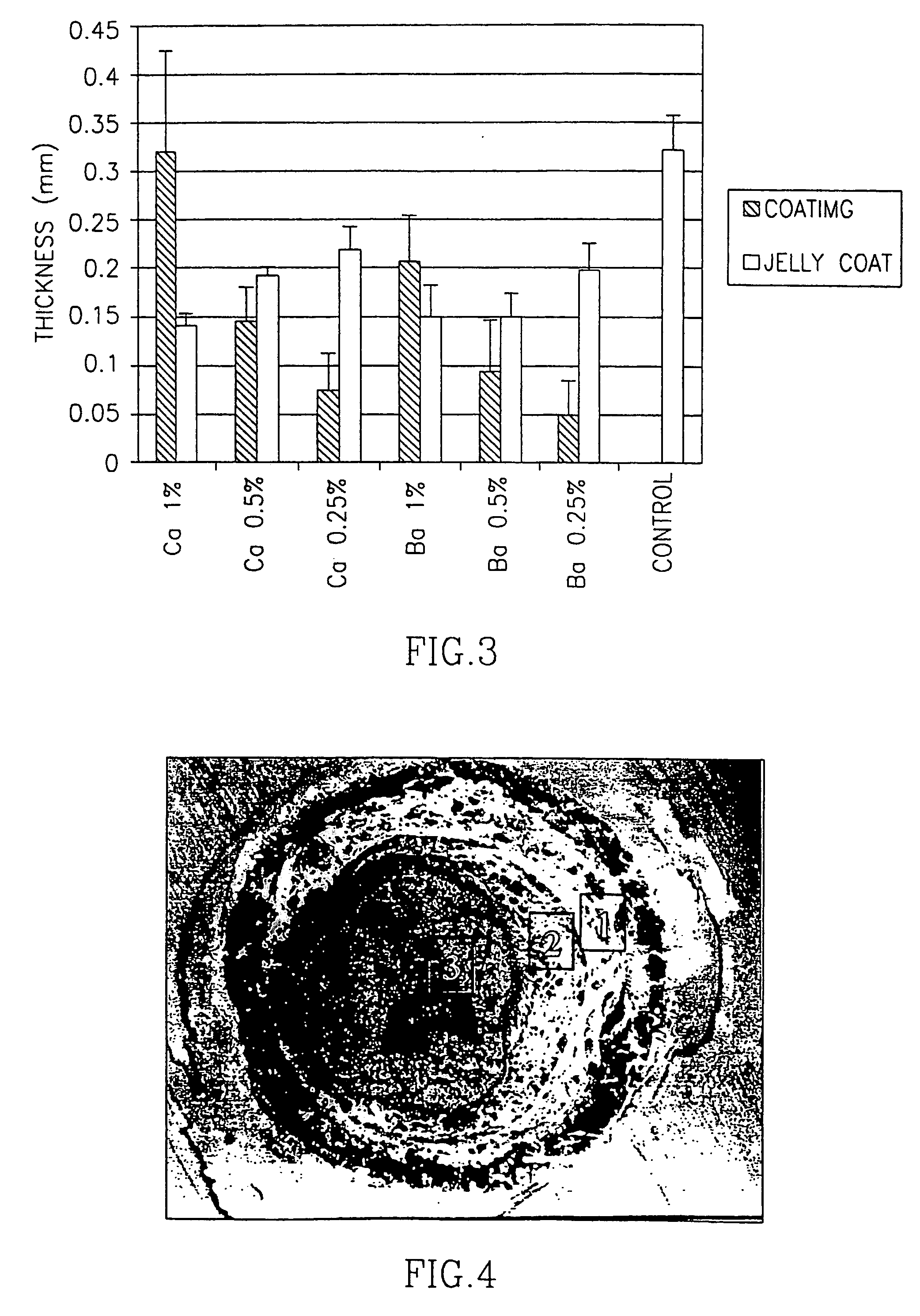
[0090] In a first set of experiments, X. laevis fertilized eggs were coated with three different types of alginate. The properties of these alginates are summarized in Table 1: they differed with respect to their molecular weights, viscosities, gel strengths and the content ratios of guluronic (G) to mannuronic (M) acid. The molecular weight, and the proportion and arrangement of M and G are expected to affect a particular alginate's behavior. The percentage of M in the alginates used for coating ranged from 29 to 35 in the alginates extracted from Laminaria hyperborea, to 61 in the alginate extracted from Macrocystic pyrifera. Each egg was covered with a thin layer of calcium- or barium- alginate gel.

TABLE 1Alginate Compositions (provided by the manufacturers)CompanyProduct NameOriginMolecular WeightViscosityGel Strength% Dry SolidsG:M RatioSigma ChemicalAlginic AcidMacrocystic Pyrifera60,000-70,00022% (cP) at aNot detected8839:61Co....

example 3

The Survival Percentage of Coated and Non-Coated X. laevis Embryos

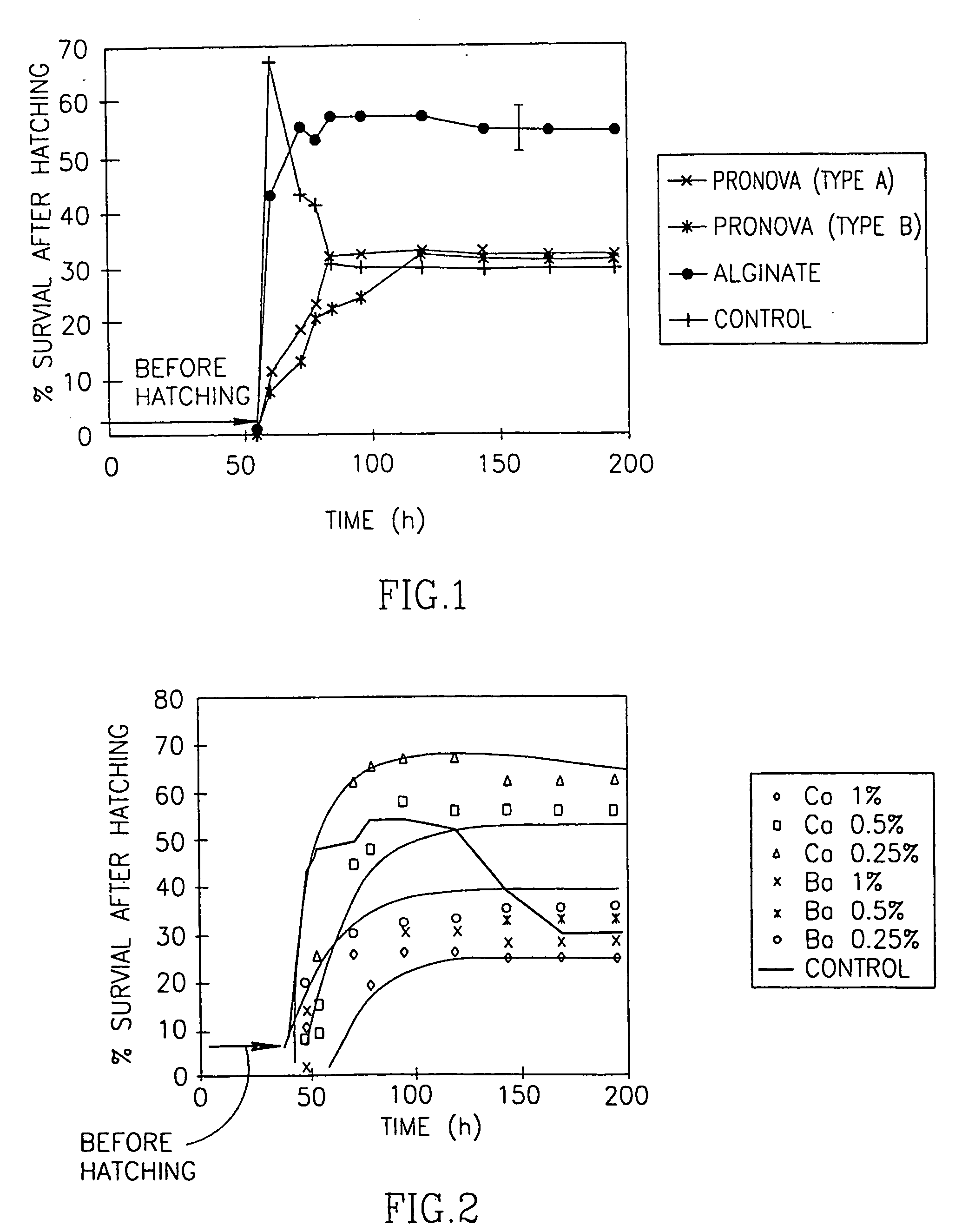
[0093] The survival of embryos vs. time under storage conditions #1 is shown in FIG. 1. The survival percentage is equivalent to the accumulated number of hatching embryos to a maximal or asymptotic survival value, and is the number of embryos left after they begin to die. The accumulated survival percentage of non-coated embryos was 4.6, 54 hours after fertilization, increasing to 66 after 60 hours (FIG. 1). Percent survival then decreased to 41 after 78 hours and reached an asymptotic value of 30 between 84 and 196 hours. Reduced survival percentages could be due to the secretion of nitrates or other substances into the medium by the developing embryos. In parallel to the survival-prospects study, embryo developmental stages were monitored (observed through a binocular lens) and compared to that of non-coated embryos (Nieuwkoop and Farber, 1994). No difference between the two was observed, implying that the coating...


PUM


Abstract

The present invention provides coated single cells or embryos having a protective micro-coating of hydrocolloid. The present invention further provides methods of coating single cells or embryos with a hydrocolloid such as an alginate, low-methoxy pectin (LMP), and carrageenans to provide a micro-coating. The coating serves as a barrier to pathogenic contamination and hazardous materials and protects against damage during freezing and thawing, thus improving survival prospects.

Description

RELATED APPLICATIONS [0001] This application is a continuation-in-part of U.S. Ser. No. 09 / 856,423 filed May 21, 2001, the specification of which is incorporated by reference herein.FIELD OF THE INVENTION [0002] The present invention relates to single cells or embryos having a protective micro-coating of hydrocolloid. In particular, the present invention relates to hydrocolloid coating of individual cells or embryos, the hydrocolloid coating being preferably less than 20% of the diameter of the cell. BACKGROUND OF THE INVENTION [0003] There is a long-felt need in biotechnology and industry for methods of protection of individual cells (e.g. stem cells or gametes, especially eggs) and embryos against pathogenic contamination, hazardous materials, and damage caused by ice crystal formation during a freezing process. Although major efforts have been made to achieve this goal, a comprehensive solution to this problem is still needed in the art. Methods of Coating and Entrapping [0004] ...

Claims


Application Information

Patent Timeline
IPC IPC(8): A01N1/02C12N11/04
CPCC12N11/04
Inventor NUSSINOVITCH, AMOSKAMPF, NIR
Owner YISSUM RES DEV CO OF THE HEBREW UNIV OF JERUSALEM LTD
Why Eureka
Try Eureka