Method of counting microorganisms or cells

a microorganism or cell technology, applied in the field of counting microorganisms or cells, can solve the problems of affecting the processing accuracy of the process, the difficulty of culture of many viable bacteria, and the inability to detect bacteria by a conventional culture method, etc., and achieve the effect of processing more accurately

Inactive Publication Date: 2006-04-06
FUJI ELECTRIC SYST CO LTD
View PDF30 Cites 13 Cited by
  • Summary
  • Abstract
  • Description
  • Claims
  • Application Information

AI Technical Summary

Benefits of technology

[0031] Preferably, the adhesive layer has a thickness of 5 to 100 μm from the viewpoint of adhesion and follow-up properties to the surface to be detected, and capture of the microorganisms. When the fluorescent image of the microorganisms or the cells is obtained, smoothness (convexoconcave difference) on the surface of the adhesive layer is within a depth of focus of the image obtaining means, and preferably and practically 20 μm or less. When the smoothness is 20 μm or less, the fluorescent image obtaining means can match the focuses more precisely, whereby the image can be processed more accurately. The smoothness can be determined by observing the cross-section of the adhesive sheet using a surface roughness tester or an electron microscope, and measuring a mean height from the summit of a convex part to the lowest of a concave part on the surface of the adhesive agent.

Problems solved by technology

Under a natural environment, many viable bacteria are difficult to be cultured.
Therefore, the bacteria may not be detected by a conventional culture method.
However, in the method of detecting the bacteria with fluorescence, when fluorescent contaminants coexist in the sample, the contaminants are false detected, resulting in a counting error.
Although the microorganisms or the cells labeled with the fluorescent reagent are more easily discolored than the fluorescent contaminants contained in the sample, fluorescent properties of the contaminants cannot be controlled.
If the fluorescent contaminants are discolored together with the microorganisms or the cells that are subjected to fluorescent labeling, measurement errors might be induced.
It is therefore not possible to count the number of the microorganisms or the cells with high accuracy.
Once the condition of the sample change, it is difficult to accurately count the number of the microorganisms or the cells.
In general, since a field of view of a microscope is small, a wide scan is needed and a long measurement time is unavoidably required.
It is thus time consuming and cumbersome.

Method used


Image

  • Method of counting microorganisms or cells
  • Method of counting microorganisms or cells
  • Method of counting microorganisms or cells

Examples


example 1

Method of Counting by Determining a Difference in the Number of Luminous Points

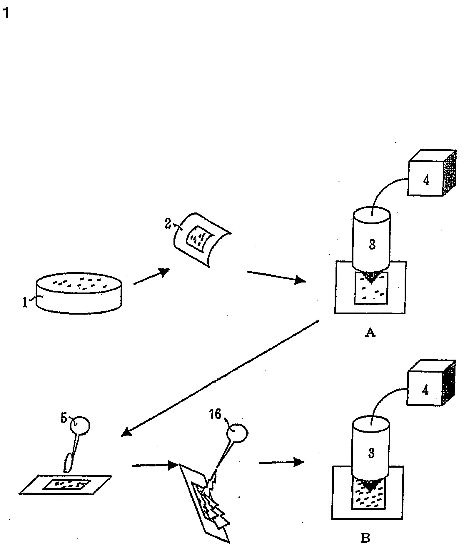
[0052] Referring to FIG. 1, the embodiment mainly according to claims 1, 5 and 6 will be described below. This embodiment relates to a method of counting the number of bacteria contained in a solid sample by utilizing a difference between luminous points in the first and second images.

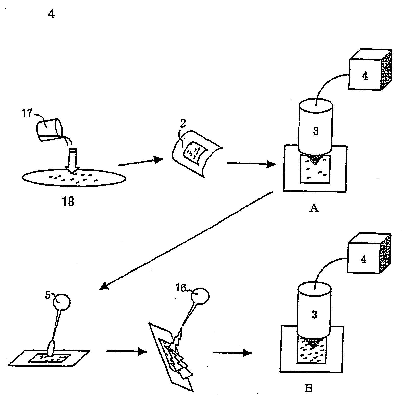
[0053] First, a solid sample 1 containing bacteria and contaminants is captured on the above-mentioned adhesive sheet 2. If the adhesive sheet is not used, the bacteria are generally captured by wiping or stomaching to deploy them into sterilizaed water. The use of the adhesive sheet simplifies the sampling operation.

[0054] Then, a means for obtaining a fluorescent image 3 is used to obtain a fluorescent image (first image) of the sample on the adhesive sheet containing the bacteria. The obtained image is image-processed at an image and arithmetic processing unit 4 to determine fluorescent luminous points A in the image. ...

example 2

Method of Counting by Utilizing a Differential Image

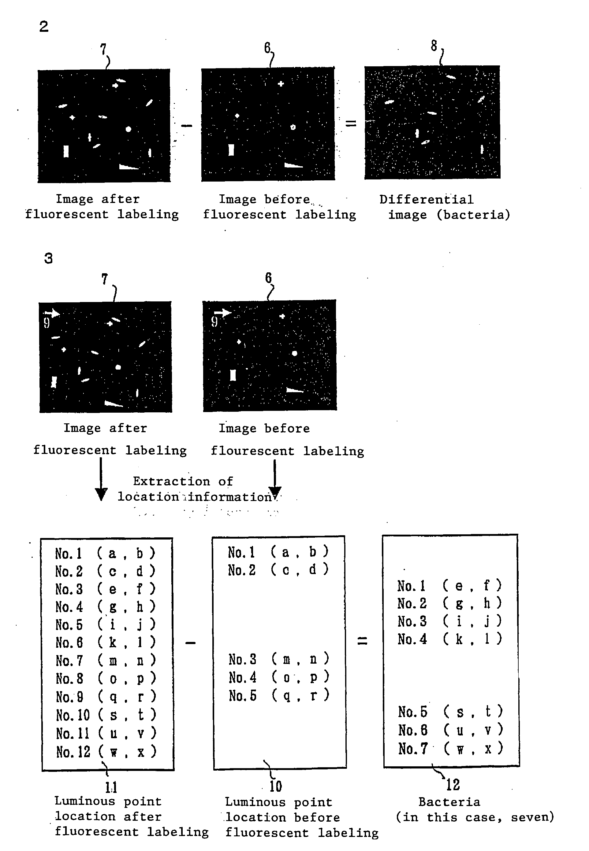
[0068] Referring to FIG. 2, the embodiment according to claim 2 will be described below. This embodiment relates to a method of counting the number of bacteria by utilizing a differential image between the first and second images.

[0069] First, a sample containing the bacteria is captured on the above-mentioned adhesive sheet similar to embodiment 1. Next, a fluorescent image of the sample containing the bacteria (a first image 6 shown in a central part of FIG. 2) on the adhesive sheet is obtained. Then, a fluorescent labeling reagent is applied over the adhesive sheet to fluorescent-label the bacteria. After the fluorescent labeling reagent that is not taken to the bacteria is cleaned with a cleaning liquid, a fluorescent image of the sample (a second image 7 shown in a left part of FIG. 2) on the adhesive sheet is obtained. A differential image 8 shown in a right part of FIG. 2 is obtained from the second image 7 and the first i...

example 3

Method of Counting by Utilizing Location Information

[0078] Referring to FIG. 3, the embodiment according to claim 3 will be described below. This embodiment relates to a method of counting the number of bacteria by utilizing a location information of luminous points.

[0079] Similar to Examples 1 and 2, the sample containing the bacteria is captured on the above-mentioned adhesive sheet. Then, a fluorescent image (first image 6) of the sample containing the bacteria on the adhesive sheet is obtained. When the image is obtained, a reference point 9 (shown in the drawing as a white arrow) is used to find the location of the sample on the adhesive sheet and to recognize the location information 10 of the luminous points in the first image. A fluorescent labeling reagent is applied over the adhesive sheet to fluorescent-labeling the bacteria. Thereafter, a fluorescent image (second image 7) of the sample containing the bacteria on the adhesive sheet is obtained. Similar to the first ima...


PUM

PropertyMeasurementUnit
thicknessaaaaaaaaaa
smoothnessaaaaaaaaaa
thicknessaaaaaaaaaa

Abstract

A method of counting microorganisms or cells in a sample by labeling the microorganisms or cells with a fluorescent labeling reagent, which comprises contacting the sample containing the microorganisms or cells with an adhesive sheet having an adhesive layer laminated on at least one surface of a substrate to capture the sample; before fluorescent-labeling the microorganisms or cells, obtaining a first fluorescent image of the sample; after fluorescent-labeling the microorganisms or the cells, obtaining a second fluorescent image of the sample, and then determining the difference in the number of luminous points between the first and second images, or determining a differential image between the first and second images and determining the number of luminous points in this differential image, or determining the number of luminous points among the luminous points in the second image located outside non-sensitive regions attached to the individual luminous points in the first image.

Description

TECHNICAL FIELD [0001] The present invention relates to a method of counting microorganisms or cells in a sample by labeling the microorganisms or the cells with a fluorescent labeling reagent, and obtaining a fluorescent image. BACKGROUND ART [0002] Detection of microorganisms in samples, and tissues and cells of animals and plants is an extremely important matter in an industrial view point in order to confirm sterilization and find abnormality of viable cells. For convenience, the following description is mainly based on bacteria. [0003] Under a natural environment, many viable bacteria are difficult to be cultured. The bacteria do not form colonies on a general agar plate medium and often do not grow on a liquid culture medium. Therefore, the bacteria may not be detected by a conventional culture method. [0004] To solve the problem, fluorescein diacetate (FDA), carboxy fluorescein diacetate (CFDA), 5-cyano-2,3-ditolyl tetrazolium chloride (CTC) and the like that are metabolized ...

Claims


Application Information

Patent Timeline
Patent Type & Authority Applications(United States)
IPC IPC(8): C12Q1/00C12Q1/04C12M1/34G01N21/64
CPCC12Q1/04G01N21/6428G01N21/6456G01N2021/6439
Inventor NODA, NAOHIROHIRAOKA, MUTSUHISATAKAHASHI, KAZUHITOMARUYAMA, KOJISAIKA, TAKESHITANAKA, YASUNOBUNASU, MASAOYAMAGUCHI, NOBUYASU
Owner FUJI ELECTRIC SYST CO LTD
Why Eureka
Try Eureka