
Crystals of cytochrome P450 2C9, structures thereof and their use

a technology of cytochrome p450 and crystallization protein, applied in the field of crystallization protein 2c9, their structure and their use, can solve the problems of complex structure, chancy and difficult process without clear expectation of success, and the crystallisation of protein molecules from solution is the major obstacle, and the balance between specific and non-specific interactions is delica

Inactive Publication Date: 2006-06-01
ASTEX THERAPEUTICS LTD
View PDF11 Cites 6 Cited by
  • Summary
  • Abstract
  • Description
  • Claims
  • Application Information

AI Technical Summary

Benefits of technology

The patent text describes a method for improving the crystallization of cytochrome P450 2C9, a membrane-associated protein. The method involves removing the N-terminal membrane anchor domain and replacing it with a short hydrophilic peptide. This modification prevents the protein from associating with membranes and promotes crystallization. The text also describes the use of a four histidine tag for purification in high salt buffers. The mutation of the protein was performed using standard recombinant DNA techniques. The technical effect of this patent is to provide a method for improving the crystallization of cytochrome P450 2C9, which has been difficult to achieve using traditional methods.

Problems solved by technology

Another level of complication results from the fact that these enzymes exhibit different tissue distributions and polymorphisms between individuals and ethnic populations
One of the greatest problems in drug discovery is the prediction of the role of cytochrome P450s on the metabolism or modification of drug leads.
It is well-known in the art of protein chemistry, that crystallising a protein is a chancy and difficult process without any clear expectation of success.
It is commonly held that crystallisation of protein molecules from solution is the major obstacle in the process of determining protein structures.
The reasons for this are many; proteins are complex molecules, and the delicate balance involving specific and non-specific interactions with other protein molecules and small molecules in solution, is difficult to predict.
Simply supersaturating the protein to bring it out of solution may not work, the result would, in most cases, be an amorphous precipitate.
Many kits are available (e.g. from Hampton Research), which attempt to cover as many parameters in crystallisation space as possible, but in many cases these are just a starting point to optimise crystalline precipitates and crystals which are unsuitable for diffraction analysis.
Even so, crystallisation of proteins is often regarded as a time-consuming process, whereby subsequent experiments build on observations of past trials.
In cases where protein crystals are obtained, these are not necessarily always suitable for diffraction analysis; they may be limited in resolution, and it may subsequently be difficult to improve them to the point at which they will diffract to the resolution required for analysis.
It may be due to intrinsic mobility of the protein within the crystal, which can be difficult to overcome, even with other crystal forms.
It may be due to high solvent content within the crystal, which consequently results in weak scattering.
Alternatively, it could be due to defects within the crystal lattice which mean that the diffracted x-rays will not be completely in phase from unit to unit within the lattice.
Any one of these or a combination of these could mean that the crystals are not suitable for structure determination.
It is often hard to predict how a protein could be re-engineered in such a manner as to improve crystallisability.
Our understanding of crystallisation mechanisms are still incomplete and the factors of protein structure which are involved in crystallisation are poorly understood.

Method used


Image

  • Crystals of cytochrome P450 2C9, structures thereof and their use
  • Crystals of cytochrome P450 2C9, structures thereof and their use
  • Crystals of cytochrome P450 2C9, structures thereof and their use

Examples


example 1

Co-Crystallisation of 2C9-FGloop K206E with S-Warfarin

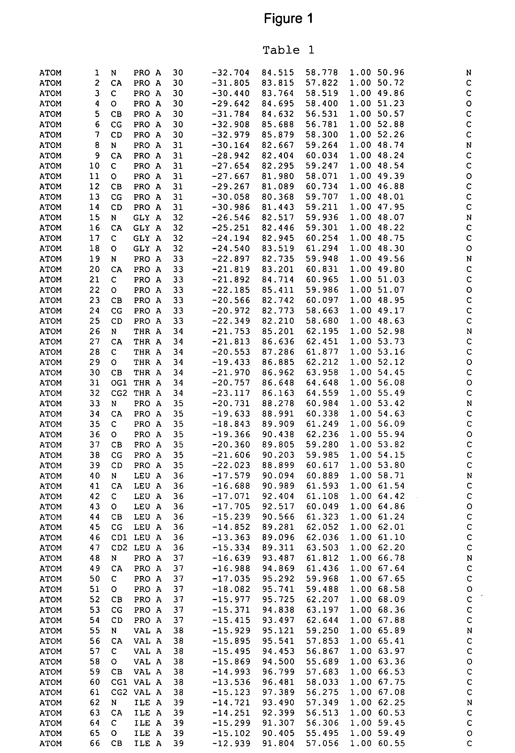
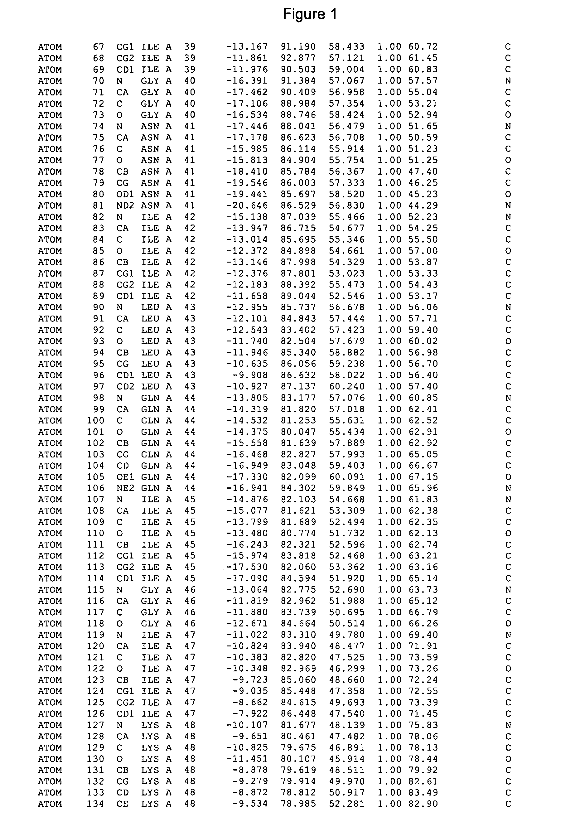
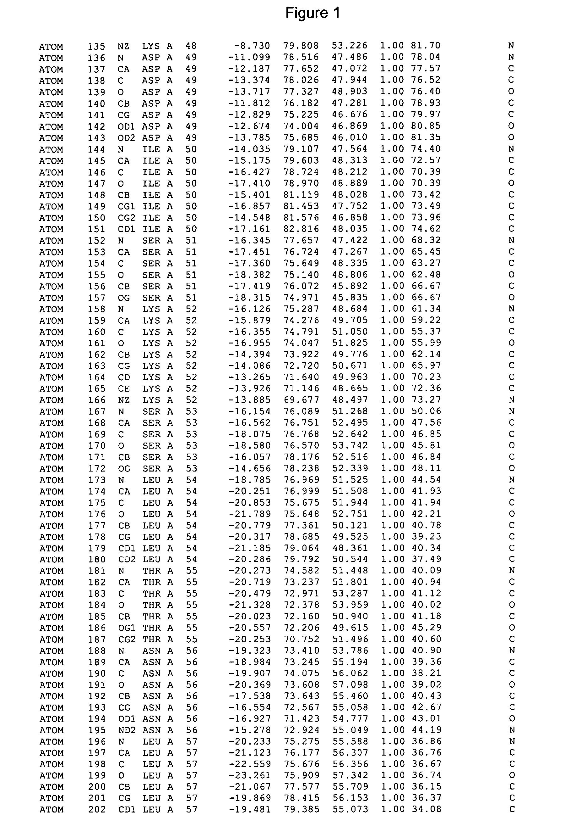
[0368] CYP 2C9 catalyses the 6- and 7-hydroyxiation of the active enantiomer of warfarin, S-warfarin, to inactive metabolites. To explore the molecular basis of drug recognition we have determined the crystal structure of CYP 2C9 complexed with S-warfarin.

[0369] 2C9-FGloop K206E was produced in E. coli as described in Annex 1, the contents of which correspond to Example 1, 2, and 8 of WO03 / 035693. To establish the effect of the truncation and mutagenesis, a comparison of the activity and specificity of the protein was performed, and it was confirmed that the 6' and 7' hydroxylation of warfarin remained unchanged compared to wild-type 2C9.

[0370] Crystals were obtained by the hanging drop vapour diffusion method, using a 1:1 ratio of protein at 40 mg / ml in a solution of 10 mM potassium phosphate, pH 7.4, 0.5M potassium chloride, 20% (v / v) glycerol, 1 mM EDTA, 2 mM DTT against a crystallisation well solution of 0.1M Tris, pH 8.4, 15...

example 2

Relevance of the S-Warfarin Remote Binding Site for Drug Metabolism in Human Cytochrome P450 2C9

[0393] In this example the relevance of the remote warfarin biding site identified above is illustrated.

[0394] Cytochrome P450s 2C9trunc (1003, SEQ ID N04) and 2C9-FGloop K206E (1155, SEQ ID NO:2) metabolize the biologically active enantiomer of warfarin, S-warfarin one of the most widely prescribed oral anticoagulant, to the 6- and 7-hydroxyl metabolites to terminate the action of the drug. The structure of 2C9-FGloop K206E complexed with S-warfarin determined at 2.6 A resolution has revealed a new binding mode of warfarin distant from the heam, at the entry of the substrate channel, near the B'-C and F-G regions. To validate this remote binding pocket and to address its physiological relevance in drug metabolism in 2C9, residues L102, A103, L208 and N217, located in this remote binding pocket at .about.3.5 A from the S-warfarin molecule, have been mutated by site directed mutagenesis an...

example 3

Back-Soaking of 2C9FGloop K206E -Warfarin Crystals

[0413] Generation of the 2C9-S-Warfarin Complex Crystals.

[0414] Co-crystals of 2C9 construct 1155 with S-warfarin are generated in a similar way to the generation of apo crystals. To order to obtain suitably large, well formed crystals it remains necessary to set up a limited grid screen around a known crystallization condition. This is typically achieved by setting up crystallizations using the conditions 0.1 M Tris pH 8-8.8, 15-30% PEG 400, 5% PEG 8000, 10% Glycerol

[0415] It may prove necessary to vary some of the crystallization variables (e.g. buffer pH, precipitant concentration) further than in the screen described above. A crystallization tray is pipetted out, with each crystallization well containing 1 ml of the above solutions. A stock solution of 0.2M S-warfarin is generated by dissolving S-warfarin in 40% DMSO, 60% ethanol. 19 .mu.l of the first well solution is then removed, placed in an eppendorf and 1 .mu.l of the 0.2M ...


PUM

PropertyMeasurementUnit
concentrationaaaaaaaaaa
pHaaaaaaaaaa
pHaaaaaaaaaa

Abstract

The present invention provides co-crystals of cytochrome P450 2C9 proteins and a ligand such as warfarin which has been crystallised to provide a high resolution structure. The structure may be used for homology modelling of other cytochrome P450 structures such as 2C8, 2C18 and 2C19, and for analysis of the interaction of ligands with P450.

Description

[0001] This application is a continuation of PCT / GB2004 / 001864, which designated the U.S. and was filed Apr. 30, 2004 (pending); the present application is also a continuation-in-part of U.S. application Ser. No. 10 / 426,058, filed Apr. 30, 2003 (pending), the present application is also a continuation-in-part of U.S. application Ser. No. 10 / 280,137, filed Oct. 25, 2002 (pending), and U.S. application Ser. No. 10 / 280,137 claims benefit of priority of U.S. Provisional Application No. 60 / 330,585, filed Oct. 25, 2001; U.S. Provisional Application No. 60 / 339,421, filed Dec. 14, 2001; U.S. Provisional Application No. 60 / 341,267, filed Dec. 20, 2001; and U.S. Provisional Application No. 60 / 396,588, filed Jul. 18, 2002; the entire contents of each of the above-identified applications being incorporated herein by reference.[0002] The present invention relates to a co-crystals of human cytochrome P450 protein 2C9 and a ligand such as S-warfarin, methods of production of co-crystals of 2C9, an...

Claims


Application Information

Patent Timeline
Patent Type & Authority Applications(United States)
IPC IPC(8): G06F19/00C12N9/02
CPCC07K14/80
Inventor WILLIAMS, PAMELA ANNCOSME, JOSE MARIEVINKOVIC, DIJANA MATAKMURRAY, CHRISTOPHER WILLIAMJHOTI, HARREN
Owner ASTEX THERAPEUTICS LTD
Learn More