Fibrils
a technology of fibrils and fibrils, applied in the field of fibrils, can solve problems such as cell death and tissue malfunction, and achieve the effect of speeding up the process
- Summary
- Abstract
- Description
- Claims
- Application Information
AI Technical Summary
Benefits of technology
Problems solved by technology
Method used
Image

Examples
example 1
Microscopy and Image Classification
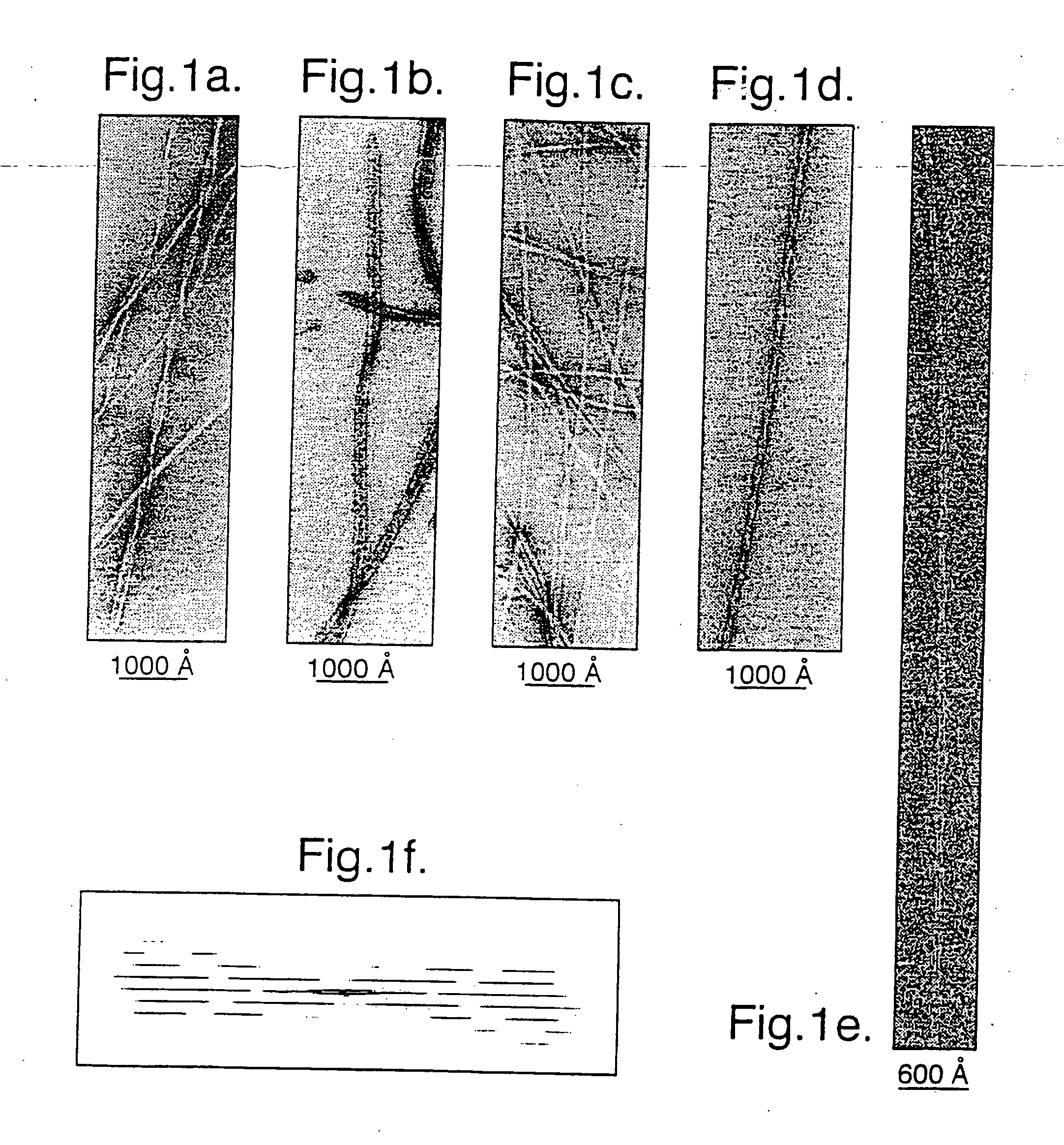
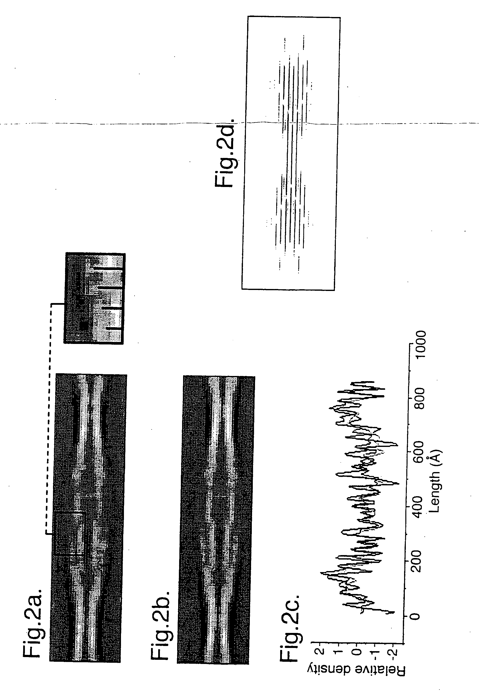
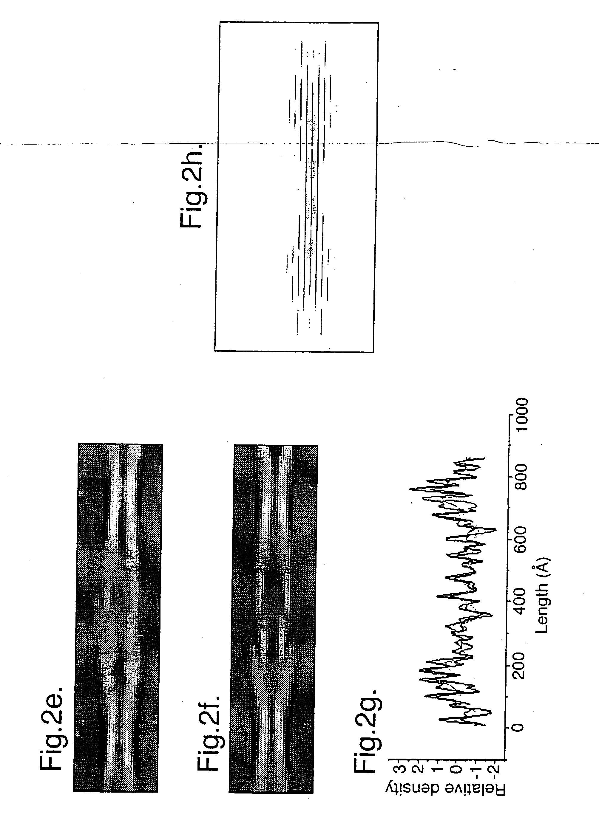
[0051] Samples of twisted fibrils of the PI3-kinase SH3 domain formed after several months incubation at pH 2 (J. I. Guijarro et al (1998) Proc. Natl. Acad. Sci. USA. 95, 4224-4228) were vitrified on holey carbon grids, and low electron dose images were recorded at 120 kV and 1.3-1.5 μm underfocus on a JOEL 1200 EX microscope with an Oxford Instruments cryotransfer stage at 30,000×. Films were digitised on a Leafscan 45 linear CCD scanner (Ilford Ltd, Cheshire, UK) at a spacing of 10 μm, and interpolated to 0.67 nm / pixel for processing. Calculated diffraction patterns (FIG. 1f) were obtained by straightening fibres with Phoelix software, but the axial resolution was severely limited in the pitch, which ranged from 54.5 to 66 nm. In order to avoid resolution loss due to non-linear interpolation, digitised fibres were cut into individual repeats and treated as single particles. 890-cut-out repeats were iteratively aligned and sorted into classes by...
example 2
Example 2 (i)
[0060] Muscle acylphosphatase was purified as previously reported (A. Modesti et al. (1995) Protein Express Purif. 6, 799) and incubated at a concentration of 0.375 mg / ml (34 μM) in 25% v / v trifluoroethanol (TFE), acetate buffer, pH 5.5 at 25° C. under constant stirring. Aliquots were withdrawn at regular time intervals for electron microscopy and spectroscopic analysis. Circular dichroism spectra were acquired directly by means of a Jasco J-720 spectropolarimeter and cuvettes of 1 mm path length. Electron micrographs were acquired by a JEM 1010 transmission electron microscope at 80 kV excitation voltage. A 3 μL sample of protein solution was placed and dried for five minutes on a Formvar and carbon-coated grid. The sample was then stained with 3 μL 1% phosphotungstic acid solution and observed at magnifications of 25-100k.
Example 2 (ii)
[0061] Infrared spectra were acquired using BaF2 windows of 50 μm path length.
example 2 (
iii)
[0062] Thioflavin T and Congo Red assays were performed according to Le Vine III (H. Le Vine III (1995) Amyloid: Int. J. Exp. Clin. Invest. 2,1.) and Klunk (W. E. Klunk et al. (1989) J. Histochem. Cytochem. 37, 1293), respectively. For Congo Red birefringence experiments aliquots of protein were air dried onto glass slides. The resulting films were stained with a saturated solution of Congo red and sodium chloride, corrected to pH 10.0 with 1% sodium hydroxide. The stained slides were examined by an optical microscope between crossed polarizers.
[0063] There is increasing evidence that amyloids develop not directly from the native and functional conformation of the protein, but from an amyloidogenic precursor bearing scant resemblance with the conformation of the native protein and identifiable in a denatured conformation containing a certain level of residual structure. This conformation is often referred to as amyloidogenic intermediate.
[0064] Muscle acylphosphatase is a prot...
PUM
| Property | Measurement | Unit |
|---|---|---|
| Temperature | aaaaa | aaaaa |
| Temperature | aaaaa | aaaaa |
| Temperature | aaaaa | aaaaa |
Abstract
Description
Claims
Application Information
 - R&D
- Intellectual Property
- Life Sciences
- Materials
- Tech Scout
- Unparalleled Data Quality
- Higher Quality Content
- 60% Fewer Hallucinations
Browse by: Latest US Patents, China's latest patents, Technical Efficacy Thesaurus, Application Domain, Technology Topic, Popular Technical Reports.
© 2025 PatSnap. All rights reserved.Legal|Privacy policy|Modern Slavery Act Transparency Statement|Sitemap|About US| Contact US: help@patsnap.com