
Methods for isolating and characterizing endogenous mRNA-protein (mRNP) complexes

a technology of mrna-protein complexes and complexes, applied in the field of posttranscriptional regulation and methods of profiling gene expression, can solve the problems of inability to report on in vitro applications, and inability to independently assess each process en mass

Inactive Publication Date: 2006-07-06
RIBONOMICS
View PDF29 Cites 21 Cited by
  • Summary
  • Abstract
  • Description
  • Claims
  • Application Information

AI Technical Summary

Benefits of technology

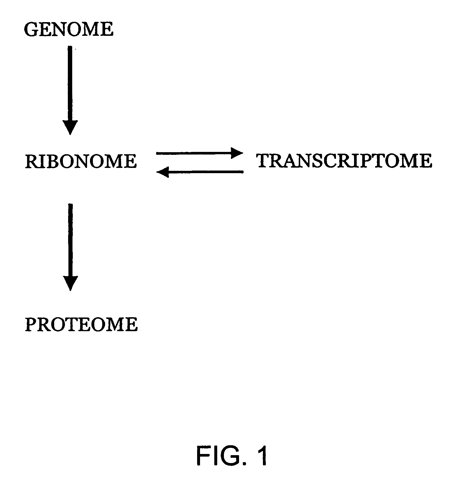
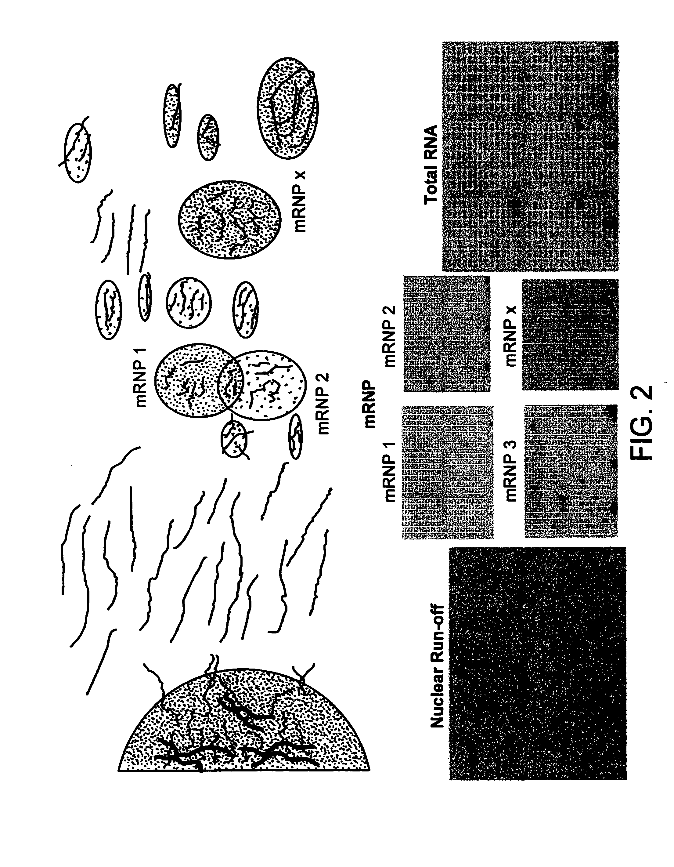
[0011] The present invention relates to a new, in vivo approach for the determination of gene expression that utilizes the flow of genetic information through messenger RNA clusters or subsets. Recently, the practice of examining multiple macromolecular events simultaneously and in parallel with the goal of organizing such information computationally has taken the designation “-ome.” Thus, the genome identifies all of the genes of a cell, while the transcriptome is defined as the messenger RNA complement of the genome and the proteome is defined as the protein complement of the genome (see FIG. 1). The present inventors have defined several physically organized subsets of the transcriptome and defined them as dynamic units of the “ribonome”. As described herein, the ribonome consists of a plurality of distinct subsets of messenger RNAs (mRNAs) that are clustered in the cell due to their association with RNA-binding proteins (e.g., regulatory RNA-binding proteins). By identifying the mRNA components of a cellular ribonome, the cellular transcriptome can be broken down into a series of subprofiles that together can be used to define the gene expression state of a cell or tissue (see FIG. 2). In combination with, for example, high throughput approaches and by multiplexing RNA processing assays, the present inventive methods provide the ability to determine the changes that occur in multiple gene transcripts simultaneously.
[0018] In certain embodiments, therefore, the present invention advantageously allows the artisan to identify, monitor, and quantitate mature gene transcripts en masse in order to determine their localization, activity, stability, and translation into protein components of living cells. The methods described herein advantageously provide a novel approach to functional genomics by providing methods of isolating endogenous messenger-RNA binding proteins, and methods of identifying the subset of cellular mRNAs contained in mRNP-complexes, using microarrays or other known procedures. In preferred embodiments, the inventive method provides a basis for investigating and determining functional mRNA networks during growth and differentiation cycles by using mRNA-binding proteins and other mRNP-associated factors to define mRNA subsets.
[0020] Gene expression profiles will also vary between differing cell types present in a complex tissue, such as a tumor. Some mRNA binding proteins are present only in certain tumor cells, and a tumor may comprise more than one cell type. Gene expression profiling for each cell type within a tumor or tissue may be carried out by making an extract of the tissue and immunoprecipitating cell-type specific components of mRNP complexes (e.g., RNA-binding proteins that are attached to mRNA) directly from the extract (i.e., in vivo). The immunoprecipitated pellets will contain mRNAs that are only present in the same cells that contain the attached or associated component. Thus, in certain embodiments, the inventive methods may be used to characterize and distinguish the gene expression profiles of a plurality of cell types, which cell types may co-exist in the same complex tissue. This can allow the tumor cells to be profiled in whole tumor extracts without having to analyze mRNA in, for example, the non-tumor stromal cells and blood cells that surround tumor cells. The results of such characterization may be useful in determining, for example, the proper course of treatment for a patient suffering with a tumor, when the choice of treatment depends of the kind of tissue (e.g., endothelial vascular tissue) present in a tumor.

Problems solved by technology

Although the sum total of these regulatory processes in each cell accounts for its unique expression profile, few methods are available to independently assess each process en masse.
This is very laborious, as only a small amount of cellular material is recovered and the purity as well as the quality of the cellular material is compromised.
However, because of post-transcriptional events affecting mRNA stability and translation, the expression levels of many cellular proteins do not directly correlate with steady-state levels of mRNAs (Gygi et al.
However, these reports are limited to in vitro applications, and do not describe in vivo methods for partitioning RNA into structural or functional subsets using RNA binding proteins.
Although in vitro methods have been used to determine protein-RNA interactions, their use has certain limitations.
Biochemical methods are generally reliable when carefully controlled, but RNA-binding can be problematic because many interactions may be of low affinity, low specificity or even artifactual.

Method used


Image

  • Methods for isolating and characterizing endogenous mRNA-protein (mRNP) complexes
  • Methods for isolating and characterizing endogenous mRNA-protein (mRNP) complexes
  • Methods for isolating and characterizing endogenous mRNA-protein (mRNP) complexes

Examples


example 1

RNase Protection in a Multiprobe System: Materials and Methods

[0078] It has previously been reported that HuB (Hel-N1) immunoprecipitation, using a g10 epitope tag, resulted in the co-immunoprecipitation of a mRNA, which once amplified by RT-PCR and sequenced, was found to encode NF-M protein (Antic, 1999, supra). In this example, the same approach is expanded to using a multiprobe RNase protection assay to rapidly optimize the immunoprecipitation of several endogenous mRNA-protein (mRNP) complexes containing different mRNA-binding proteins. In the multi-probe system, many mRNAs, from mRNP pellets, can be assayed in a single lane of polyacrylamide gel.

[0079] Cell Culture and Transformation. Murine P19 embryonal carcinoma cells were obtained from the ATCC and maintained in monolayer culture using alpha.-MEM without phenol red (Gibco-BRL 41061-029) supplemented with 7.5% Bovine Calf Serum, 2.5% Fetal Bovine Serum (Hyclone) and 100U Penicillin / Streptomycin. Cells were grown in tissue...

example 2

RNase Protection in a Multiprobe System: Experimental Results

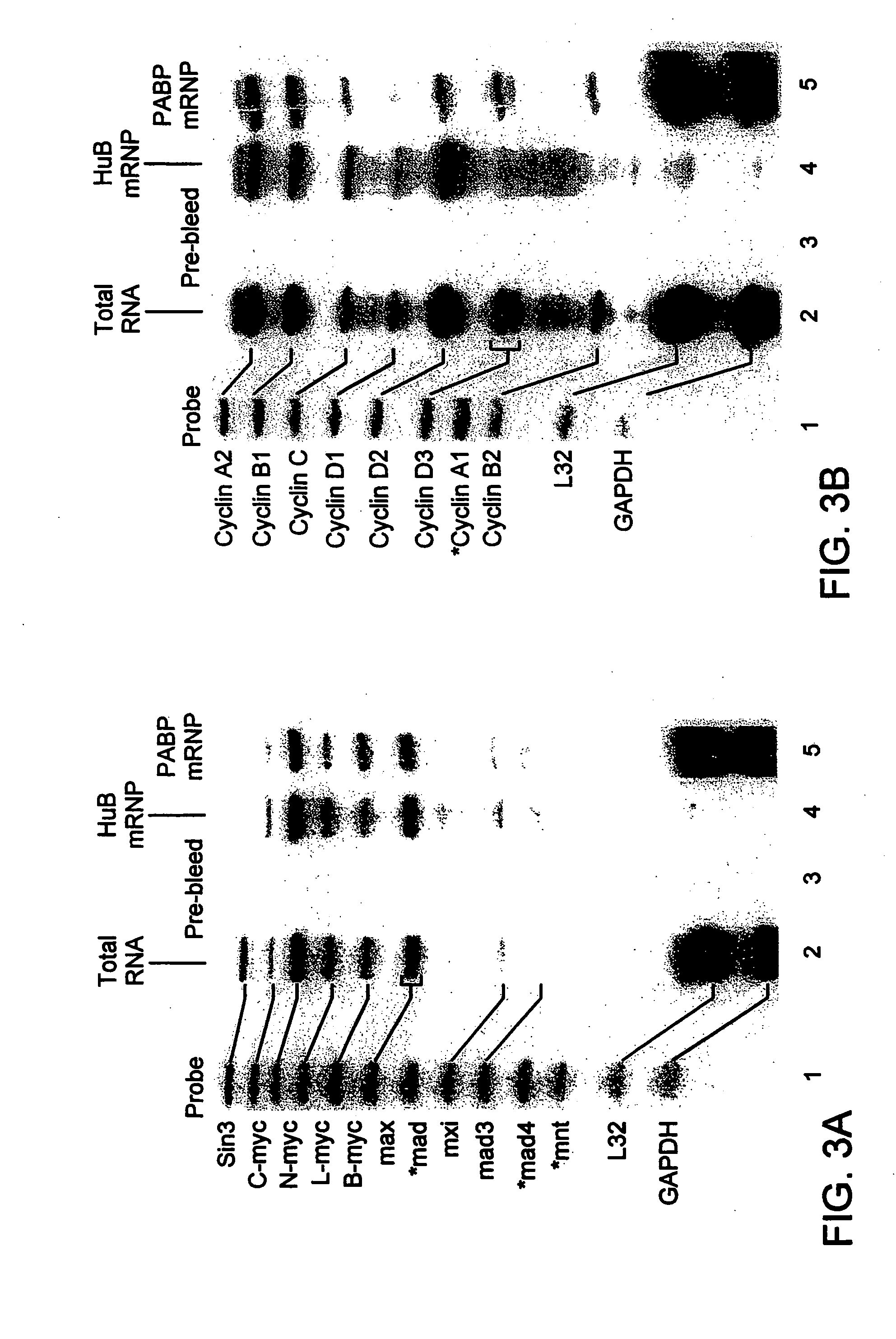
[0085]FIG. 3 shows an immunoprecipitation of HuB and Poly-A binding protein (PABP)-mRNP complexes from extracts of murine P19 cells stably transfected with g10-HuB cDNA. No mRNAs were detected in pellets immunoprecipitated with polyclonal pre-bleed rabbit sera (FIGS. 3A and 3B, lane 3), or with many other rabbit, mouse, and normal human sera tested with this assay (data not shown). The profiles of mRNAs associated with HuB mRNP complexes included n-myc, 1-myc, b-myc, max and cylins A2, B1, C, D1, and D2, but not sin3, cyclin D3, cyclin B2, L32 or GAPDH mRNAs (FIGS. 3A and 3B, lane 4). In contrast, the profiles of mRNAs extracted from PABP-mRNP complexes resembled the profiles of total RNA, but showed enriched levels of L32 and GAPDH and decreased levels of sin3 mRNA (FIGS. 3A and 3B, lane 5). It was concluded that antibodies reactive with these cellular RNA-binding proteins could be used to immunoprecipitate mRNP complexe...

example 3

Identification of mRNA Subsets Associated with RNA Binding Proteins En Masse Using cDNA Arrays: Materials and Methods

[0086] To further expand the ability to identify the mRNAs associated in endogenous mRNP complexes, this example describes the use of a cDNA array filter as a highly specific and sensitive method to detect a mRNA subset without amplification or iterative selection (FIG. 4).

[0087] Antibodies. Monoclonal anti-gene 10 (g10) antibodies were produced as previously described (see D. Tsai et al., Proc. Natl. Acad. Sci. USA, 89, 8864-8868 (1992); Gao et al. (1994) Proc. Natl. Acad. Sci. USA 91, 11207-11211; Antic et al. (1999) Genes Dev. 13, 449-461). Polyclonal sera reactive with Hu proteins were produced as previously described (Levine et al. (1993) Mol. Cell. Biol. 13, 3494-3504; Atasoy et al. (1998) J. Cell Sci. 111, 3145-3156). Antibody against 5′ cap binding protein (elF-4E) was obtained from Transduction Laboratories (San Diego, Calif.). Antibodies reactive with Poly...


PUM

PropertyMeasurementUnit
pHaaaaaaaaaa
pHaaaaaaaaaa
pHaaaaaaaaaa

Abstract

Cellular mRNA-protein (mRNP) complexes are partitioned in vivo by contacting a biological sample with at least one ligand that specifically binds at least one component of a mRNP complex. Suitable biological samples comprise at least one mRNA-protein (mRNP) complex and include cell cultures, cell extracts, and whole tissue, including tumor tissue. Ligands include antibodies that specifically bind RNA-binding or RNA-associated proteins present in the mRNP complex. The mRNP complex is separated by binding the ligand with a binding molecule specific for the ligand, where the binding molecule is attached to a solid support. The mRNP complex is collected by removing the mRNP complex from the solid support. After collecting the mRNP complex, the mRNA bound within the complex may be characterized and identified. Subsets of the total mRNA population of a cell may accordingly be characterized, and a gene expression profile of the cell obtained.

Description

RELATED APPLICATIONS [0001] This application is a continuation of U.S. patent application Ser. No. 10 / 238,306, filed on Sep. 10, 2002, which is a continuation of U.S. patent application Ser. No. 09 / 750,401, filed on Dec. 28, 2000, now issued as U.S. Pat. No. 6,635,422, which claims the benefit of U.S. Provisional Application No. 60 / 173,338, filed Dec. 28, 1999, the contents of which are hereby incorporated in their entirety.STATEMENT OF FEDERAL SUPPORT [0002] This invention was made with government support under grant number R01 CA79907 from the National Institutes of Health. The United States government has certain rights to this invention.FIELD OF THE INVENTION [0003] This invention relates generally to post-transcriptional regulation and methods of profiling gene expression. BACKGROUND OF THE INVENTION [0004] Many diseases are genetically based, and the genetic background of each individual can have a profound effect on his or her susceptibility to disease. The relatively new fie...

Claims


Application Information

Patent Timeline
Patent Type & Authority Applications(United States)
IPC IPC(8): C12Q1/68C12P21/06B01D15/08G01N33/53C07H21/02C07K1/22C12N15/09C12Q1/6809G01N33/566G01N33/68
CPCC07H21/02C12Q1/6804C12Q1/6809C12Q2600/158G01N33/5008G01N33/5023G01N33/5308G01N33/68G01N33/6803G01N33/6845C12Q2522/101
Inventor KEENE, JACKTENENBAUM, SCOTTCARSON, CRAIG
Owner RIBONOMICS
Learn More