Navigable, multi-positional and variable tissue ablation apparatus and methods

a tissue ablation and multi-position technology, applied in the field of navigation, multi-positional and variable tissue ablation apparatus and methods, can solve the problems of affecting time constants, affecting treatment procedures, and destroying tissue with some volume in the vicinity of the electrode, so as to accurately determine the location of the electrodes

Inactive Publication Date: 2006-09-07
MOWERY THOMAS M
View PDF8 Cites 186 Cited by
  • Summary
  • Abstract
  • Description
  • Claims
  • Application Information

AI Technical Summary

Benefits of technology

[0028] A spinal nerve tissue ablation apparatus includes a stylet needle with a distal end having a rounded blunt tip sufficiently sharp to penetrate tissue but sufficiently blunt to avoid impinging on bony surfaces of spinal vertebra. The apparatus includes an energy delivery device having at least a first electrode and a second electrode. Each electrode has a tissue piercing distal end and is positionable in the stylet as the stylet is advanced through tissue. The first and second electrodes are deployable with curvature from the stylet. The stylet includes rotational means for orienting the first and second electrodes and directing the extension according to the curvature of the electrodes. The stylet further includes an optical imaging module to provide continual progressive feedback of ablation surface development. The apparatus further includes an ultra-wide band radio frequency scanning device capable of accurately determining the location of the electrodes within the spinal structure.

Problems solved by technology

Of these methods, the radiofrequency (RF) and direct current heating methods involve the passage of current from an electrode to the surrounding tissue, thereby heating and destroying the tissue with some volume in the vicinity of the electrode.
Additionally, current temperature sensing methodologies are unable to account for the heat which may develop in surrounding tissues, e.g., bone, creating an additional thermal reservoir of heat, which will affect time constants and complicate treatment procedures.
The 25 gauge needle is typically not used for electrode delivery because it tends to be too flimsy to effectively penetrate percutaneous tissue.
However, a physician has great difficulty in attempting to place a single needle electrode tip adjacent to target nerves due to several factors including: (1) the geometric complexity of the spinal column, (2) the variable locations of nerves, (3) the variable and intricate paths of nerves, and, (4) differences from person to person.
Unfortunately, the structural complexity of the spine and the variable location of target nerves can sometimes limit the ability of a physician to manipulate and align a single needle in such a manner.
However, in most other spinal applications, a single needle application is more difficult to use and will not ensure adequate treatment at the target site.
In particular, without multiple needle placements, single needle electrode methodology is substantially less effective for treating sympathetic ganglia, dorsal root ganglia, medial branch nerves to facets, and peripheral nerves, among others.
The first, RF ablation, is a destructive methodology.
These repetitive patterns tend to increase patient discomfort, increase the potential for harmful needle placement and extend the time and consequently the exposure to harmful x-rays required to provide the treatment.
In particular, the treating physician, dealing with a number of patients, will normally suffer a much higher x-ray exposure from the fluoroscopy than any individual patient, thus increasing the likelihood of potentially negative future consequences for the physician.
Currently available pain management treatment systems and methods are inadequate.
Procedures can be quite uncomfortable, sporadically effective and very costly.
The high cost and limited effectiveness limits the availability of the treatment to a large portion of the population.
Additionally, it generally impedes a physician's ability to obtain approval for payment for the procedures from insurance providers.
Further, direct, real-time visualization of needle position and navigation, without dangerous x-ray exposure from fluoroscopy, is a highly desirable, but currently unavailable, technological capability.
Also, three-dimensional visualization is very desirable, but generally unavailable.

Method used


Image

  • Navigable, multi-positional and variable tissue ablation apparatus and methods
  • Navigable, multi-positional and variable tissue ablation apparatus and methods
  • Navigable, multi-positional and variable tissue ablation apparatus and methods

Examples


Embodiment Construction

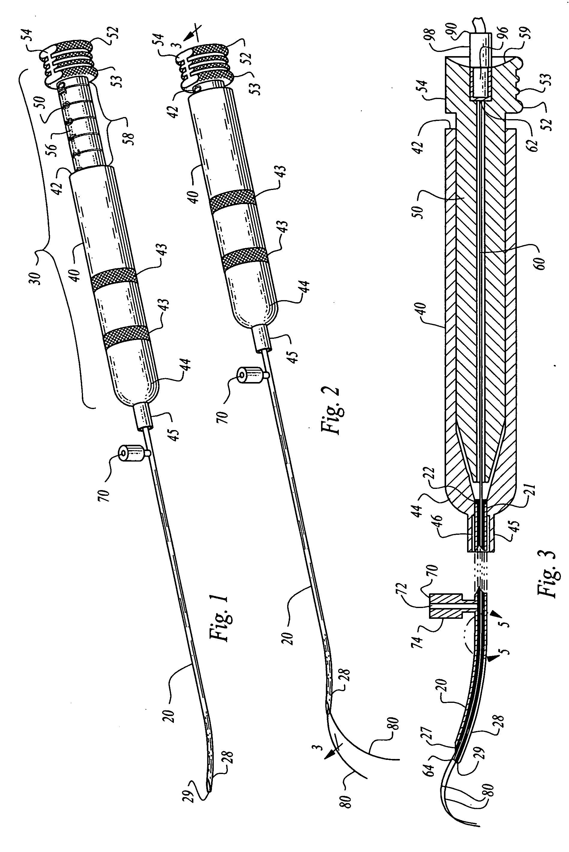
[0073] Referring to FIG. 1 and FIG. 2, a tissue ablation apparatus 10, hereinafter, the VIPER 10 includes a cylindrical tubular stylet 20. The stylet 20 is connected to a proximal control assembly 30. The control assembly 30 includes a female housing 40 and a male linear / rotational control member 50. The control member 50 is connected to a mandrel 60. The mandrel 60 is slidably and rotatably received within the stylet 20. A side infusion port 70 is disposed on a side of the stylet 20. One or more tines 80 are disposed and captured within the mandrel 40. The tines 80 are connected to the control member 50 of the control assembly 30. The control member 50 is further electrically and optically connected to an external system monitoring and control unit.

[0074] In further detail, the VIPER 10 includes a distal cylindrical elongate tubular stylet 20 fixably attached to a proximal control assembly 30. The stylet 20 is sufficiently small and made of sufficiently rigid material, such as sur...


PUM


Abstract

A spinal nerve tissue ablation apparatus includes a stylet needle with a distal end having a rounded blunt tip sufficiently sharp to penetrate tissue but sufficiently blunt to avoid impinging on bony surfaces of spinal vertebra. The apparatus includes an energy delivery device having at least a first electrode and a second electrode. Each electrode has a tissue piercing distal end and is positionable in the stylet as the stylet is advanced through tissue. The first and second electrodes are deployable with curvature from the stylet. The stylet includes rotational means for orienting the first and second electrodes and directing the extension according to the curvature of the electrodes. The stylet further includes an optical imaging module to provide continual progressive feedback of ablation surface development. The apparatus further includes an ultra-wide band radio frequency scanning device capable of accurately determining the location of the electrodes within the spinal structure.

Description

CROSS REFERENCE TO RELATED APPLICATIONS [0001] Priority is claimed to U.S. Provisional Application No. 60 / 658,112 filed Aug. 10, 2004, titled “NAVIGABLE, MULTI-POSITIONAL AND VARIABLE TISSUE ABLATION APPARATUS AND METHODS” which is referred to and incorporated herein in its entirety by this reference.FEDERALLY-SPONSORED RESEARCH [0002] Not Applicable SEQUENCE LISTING OR PROGRAM [0003] Not Applicable DESCRIPTION BACKGROUND OF THE INVENTION [0004] 1. Field of the Invention [0005] This invention relates generally to a tissue ablation apparatus and methods for ablating tissue using radiofrequency energy, and more particularly, to such an apparatus and methods capable of effectively navigating the spinal structure and using variable width, length and orientation of electrode tines to allow effective and controlled, multi-positional creation of a desired shape lesion or treatment zone. [0006] 2. Description of the Related Art [0007] Historically, numerous techniques have been used to sele...

Claims


Application Information

Patent Timeline
Patent Type & Authority Applications(United States)
IPC IPC(8): A61B18/14
CPCA61B18/1477A61B2018/00434A61B2018/0044A61B2018/143A61B2019/5217A61B2090/3614
Inventor MOWERY, THOMAS M.
Owner MOWERY THOMAS M
Why Eureka
Try Eureka